Render Target: SSR
Render Timestamp:
4/1/2025, 11:21:44 AM EDT
4/1/2025, 3:21:44 PM UTC
Commit: 461ca8d8fe5b1efd4c01fc87e5b5eb592e2d154a
XML generation date: 2025-03-07 13:10:57.578
Product last modified at: 2024-09-13T07:01:21.634Z
1% for the planet logo
PDP - Template Name: siRNA
PDP - Template ID: *******aa36529

SignalSilence® PAK2 siRNA (Human Specific) #6366

SignalSilence® PAK2 siRNA (Human Specific): Image 1
Fluorescent detection of SignalSilence® Control siRNA (Fluorescein Conjugate) #6201 in living HeLa cells 24 hours post-transfection, demonstrating nearly 100% transfection efficiency.
This product is discontinued

Inquiry Info. # 6366

Please see our recommended alternatives.
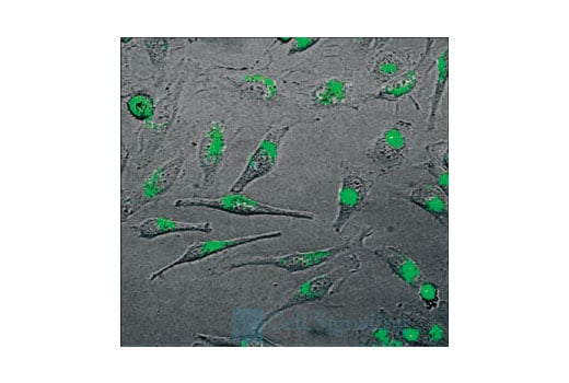
Supporting Data

REACTIVITY H
Species Cross-Reactivity Key:
  • H-Human 

Product Information

Product Usage Information

CST recommends transfection with 100nM human-specific PAK2 siRNA 48 to 72 hours prior to cell lysis. See Protocol for transfection procedure.

Storage

SignalSilence® siRNA is supplied in RNAse-free water. Aliquot and store at -20ºC.

Product Description

SignalSilence® PAK2 siRNA from Cell Signaling Technology allows the researcher to specifically inhibit PAK2 expression using RNA interference, a method in which gene expression can be selectively silenced through the delivery of double stranded RNA molecules into the cell. All SignalSilence® siRNA products are rigorously tested in-house and have been shown to reduce protein expression using total antibodies from Cell Signaling Technology.

Background

The p21-activated kinase (PAK) family of serine/threonine kinases is engaged in multiple cellular processes, including cytoskeletal reorganization, MAPK signaling, apoptotic signaling, control of phagocyte NADPH oxidase, and growth factor-induced neurite outgrowth (1,2). Several mechanisms that induce PAK activity have been reported. Binding of Rac/Cdc42 to the CRIB (or PBD) domain near the amino terminus of PAK causes autophosphorylation and conformational changes in PAK (1). Phosphorylation of PAK1 at Thr423 by PDK induces activation of PAK1 (3). Several autophosphorylation sites have been identified, including Ser199 and Ser204 of PAK1, and Ser192 and Ser197 of PAK2 (4,5). Because the autophosphorylation sites are located in the amino-terminal inhibitory domain, it has been hypothesized that modification in this region prevents the kinase from reverting to an inactive conformation (6). Research indicates that phosphorylation at Ser144 of PAK1 or Ser139 of PAK3 (located in the kinase inhibitory domain) affects kinase activity (7). Phosphorylation at Ser21 of PAK1 or Ser20 of PAK2 regulates binding with the adaptor protein Nck (8). PAK4, PAK5/7, and PAK6 have lower sequence similarity with PAK1-3 in the amino-terminal regulatory region (9). Phosphorylation at Ser474 of PAK4, a site analogous to Thr423 of PAK1, may play a pivotal role in regulating the activity and function of PAK4 (10). PAK family members are widely expressed, and often overexpressed in human cancer (11,12).

Pathways

Explore pathways related to this product.


For Research Use Only. Not For Use In Diagnostic Procedures.
Cell Signaling Technology is a trademark of Cell Signaling Technology, Inc.
SignalSilence is a registered trademark of Cell Signaling Technology, Inc.
Limited Use Label License, RNA interference: This product is licensed under European Patent 1144623 and foreign equivalents from Ribopharma AG, Kulmbach, Germany and is provided only for use in non-commercial research specifically excluding use (a) in drug discovery or drug development, including target identification or target validation, by or on behalf of a commercial entity, (b) for contract research or commercial screening services, (c) for the production or manufacture of siRNA-related products for sale, or (d) for the generation of commercial databases for sale to Third Parties. Information about licenses for these and other commercial uses is available from Ribopharma AG, Fritz-Hornschuch-Str. 9, D-95326 Kulmbach, Germany.
All other trademarks are the property of their respective owners. Visit our Trademark Information page.