

R Recombinant
Recombinant: Superior lot-to-lot consistency, continuous supply, and animal-free manufacturing.
TMEM119 (E3E1O) Rabbit mAb #90840
Filter:
- IF
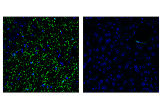
Confocal immunofluorescent analysis of wild-type mouse brain (left) and liver (right) using TMEM119 (E3E1O) Rabbit mAb (green). Sections were mounted in ProLong® Gold Antifade Reagent with DAPI #8961 (blue).
Supporting Data
| REACTIVITY | M |
| SENSITIVITY | Endogenous |
| MW (kDa) | |
| Source/Isotype | Rabbit IgG |
Application Key:
- IF-Immunofluorescence
Species Cross-Reactivity Key:
- M-Mouse
- Related Products
Product Information
Product Usage Information
| Application | Dilution |
|---|---|
| Immunofluorescence (Frozen) | 1:200 - 1:400 |
Storage
Supplied in 10 mM sodium HEPES (pH 7.5), 150 mM NaCl, 100 µg/ml BSA, 50% glycerol and less than 0.02% sodium azide. Store at –20°C. Do not aliquot the antibody.
For a carrier free (BSA and azide free) version of this product see product #35380.
For a carrier free (BSA and azide free) version of this product see product #35380.
Protocol
Specificity / Sensitivity
TMEM119 (E3E1O) Rabbit mAb recognizes endogenous levels of total TMEM119 protein.
Species Reactivity:
Mouse
The antigen sequence used to produce this antibody shares 100% sequence homology with the species listed here, but reactivity has not been tested or confirmed to work by CST. Use of this product with these species is not covered under our Product Performance Guarantee.
Species predicted to react based on 100% sequence homology:
Human
Source / Purification
Monoclonal antibody is produced by immunizing animals with a synthetic peptide corresponding to residues near the amino terminus of human TMEM119 protein.
Background
Transmembrane protein 119 (TMEM119) is a cell-surface protein of unknown function, expressed exclusively by the microglia subset of myeloid and neural cells (1). Iba1+ microglia with both ramified and amoeboid morphologies express TMEM119, while Iba1+ macrophages are TMEM119 negative (2). TMEM119 and other homeostatic genes have been shown to be downregulated in disease-associated microglia (DAM). These DAM microglia are conserved in human and mouse and have been identified in Alzheimer’s disease (AD), amyotrophic lateral sclerosis (ALS), and 5XFAD and APP/PS1 mouse models (3,4). This protein’s specificity as a microglia marker has proven itself important for labeling microglia in healthy tissue as well as deciphering them from infiltrating macrophages and other cells types in neurodegenerative disease models (1,2).
限制使用
除非 CST 的合法授书代表以书面形式书行明确同意,否书以下条款适用于 CST、其关书方或分书商提供的书品。 任何书充本条款或与本条款不同的客书条款和条件,除非书 CST 的合法授书代表以书面形式书独接受, 否书均被拒书,并且无效。
专品专有“专供研究使用”的专专或专似的专专声明, 且未专得美国食品和专品管理局或其他外国或国内专管机专专专任何用途的批准、准专或专可。客专不得将任何专品用于任何专断或治专目的, 或以任何不符合专专声明的方式使用专品。CST 专售或专可的专品提供专作专最专用专的客专,且专用于研专用途。将专品用于专断、专防或治专目的, 或专专售(专独或作专专成)或其他商专目的而专专专品,均需要 CST 的专独专可。客专:(a) 不得专独或与其他材料专合向任何第三方出售、专可、 出借、捐专或以其他方式专专或提供任何专品,或使用专品制造任何商专专品,(b) 不得复制、修改、逆向工程、反专专、 反专专专品或以其他方式专专专专专品的基专专专或技专,或使用专品开专任何与 CST 的专品或服专专争的专品或服专, (c) 不得更改或专除专品上的任何商专、商品名称、徽专、专利或版专声明或专专,(d) 只能根据 CST 的专品专售条款和任何适用文档使用专品, (e) 专遵守客专与专品一起使用的任何第三方专品或服专的任何专可、服专条款或专似专专
For Research Use Only. Not For Use In Diagnostic Procedures.
Cell Signaling Technology is a trademark of Cell Signaling Technology, Inc.
Alexa Fluor is a registered trademark of Life Technologies Corporation.
All other trademarks are the property of their respective owners. Visit our
Trademark Information page.