

R Recombinant
Recombinant: Superior lot-to-lot consistency, continuous supply, and animal-free manufacturing.
Tenascin C (E5J3B) Rabbit mAb #33352
Filter:
- WB
- IP
- IF
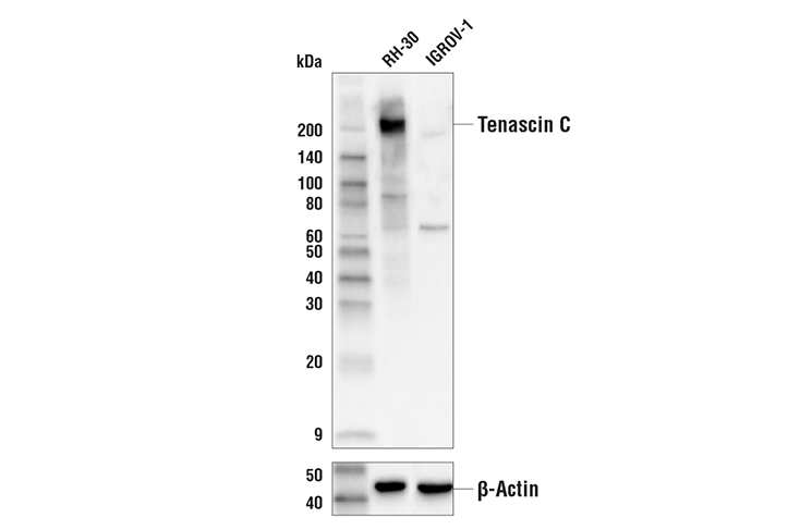
Western blot analysis of extracts from RH-30 and IGROV-1 cells using Tenascin C (E5J3B) Rabbit mAb (upper) and β-Actin (D6A8) Rabbit mAb #8457 (lower). The differential in Tenascin C expression between RH-30 and IGROV-1 cells is consistent with their reported molecular expression profiles (CCLE, portals.broadinstitute.org), confirming specificity of the antibody for Tenascin C.
Supporting Data
| REACTIVITY | H |
| SENSITIVITY | Endogenous |
| MW (kDa) | 200, 240 |
| Source/Isotype | Rabbit IgG |
Application Key:
- WB-Western Blotting
- IP-Immunoprecipitation
- IF-Immunofluorescence
Species Cross-Reactivity Key:
- H-Human
- Related Products
Product Information
Product Usage Information
| Application | Dilution |
|---|---|
| Western Blotting | 1:1000 |
| Immunoprecipitation | 1:100 |
| Immunofluorescence (Immunocytochemistry) | 1:200 - 1:800 |
Storage
Supplied in 10 mM sodium HEPES (pH 7.5), 150 mM NaCl, 100 µg/ml BSA, 50% glycerol and less than 0.02% sodium azide. Store at –20°C. Do not aliquot the antibody.
Protocol
Specificity / Sensitivity
Tenascin C (E5J3B) Rabbit mAb recognizes endogenous levels of total Tenascin C protein.
Species Reactivity:
Human
Source / Purification
Monoclonal antibody is produced by immunizing animals with recombinant protein specific to the amino terminus of human Tenascin C protein.
Background
Tenascin C is a large hexameric extracellular matrix glycoprotein that exhibits de-adhesive effects on cell-matrix interaction, enhancing cell proliferation and motility in most cell types. It is highly expressed in remodeling tissues during embryonic development and under pathological conditions in adults, and research studies have shown markedly increased expression in cancerous tissues (1,2). Tenascin C has been implicated in a variety of cellular processes relevant to atherosclerosis, including cell proliferation, migration, and apoptosis. Expression of Tenascin C is tightly controlled in adults and is upregulated in tissues undergoing wound healing (3). In development, the expression of Tenascin C is known to be associated with epithelial-mesenchymal transition (EMT) events, including gastrulation and formation of the neural crest, endocardial cushion, and secondary palate (1). Investigators have shown that Tenascin C is a key determinant of the tumor stroma and is involved in the initiation of tumorigenesis and progression to metastasis (2). Immature and mature astrocytes, radial glial cells, Schwann cells, and a subset of neurons express Tenascin C. Upon CNS trauma or exposure of neurons to excitotoxic agents, Tenascin C expression is upregulated by glial cells. Research studies have shown that Tenascin C is involved in guidance of migrating axons and neurons, synaptic plasticity, and neuronal regeneration, promoting spinal cord regeneration after injury (4).
限制使用
除非 CST 的合法授书代表以书面形式书行明确同意,否书以下条款适用于 CST、其关书方或分书商提供的书品。 任何书充本条款或与本条款不同的客书条款和条件,除非书 CST 的合法授书代表以书面形式书独接受, 否书均被拒书,并且无效。
专品专有“专供研究使用”的专专或专似的专专声明, 且未专得美国食品和专品管理局或其他外国或国内专管机专专专任何用途的批准、准专或专可。客专不得将任何专品用于任何专断或治专目的, 或以任何不符合专专声明的方式使用专品。CST 专售或专可的专品提供专作专最专用专的客专,且专用于研专用途。将专品用于专断、专防或治专目的, 或专专售(专独或作专专成)或其他商专目的而专专专品,均需要 CST 的专独专可。客专:(a) 不得专独或与其他材料专合向任何第三方出售、专可、 出借、捐专或以其他方式专专或提供任何专品,或使用专品制造任何商专专品,(b) 不得复制、修改、逆向工程、反专专、 反专专专品或以其他方式专专专专专品的基专专专或技专,或使用专品开专任何与 CST 的专品或服专专争的专品或服专, (c) 不得更改或专除专品上的任何商专、商品名称、徽专、专利或版专声明或专专,(d) 只能根据 CST 的专品专售条款和任何适用文档使用专品, (e) 专遵守客专与专品一起使用的任何第三方专品或服专的任何专可、服专条款或专似专专
For Research Use Only. Not For Use In Diagnostic Procedures.
Cell Signaling Technology is a trademark of Cell Signaling Technology, Inc.
XP is a registered trademark of Cell Signaling Technology, Inc.
All other trademarks are the property of their respective owners. Visit our
Trademark Information page.