

R Recombinant
Recombinant: Superior lot-to-lot consistency, continuous supply, and animal-free manufacturing.
S100B (E7C3A) Rabbit mAb #90393
Filter:
- WB
- IHC
- IF
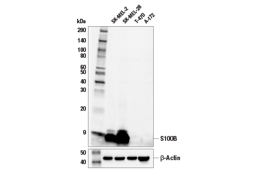
Western blot analysis of extracts from various cell lines using S100B (E7C3A) Rabbit mAb (upper) and β-Actin (D6A8) Rabbit mAb #8457 (lower).
Supporting Data
| REACTIVITY | H M R |
| SENSITIVITY | Endogenous |
| MW (kDa) | 10 |
| Source/Isotype | Rabbit IgG |
Application Key:
- WB-Western Blotting
- IHC-Immunohistochemistry
- IF-Immunofluorescence
Species Cross-Reactivity Key:
- H-Human
- M-Mouse
- R-Rat
- Related Products
Product Information
Product Usage Information
| Application | Dilution |
|---|---|
| Western Blotting | 1:1000 |
| Simple Western™ | 1:10 - 1:50 |
| Immunohistochemistry (Paraffin) | 1:100 - 1:400 |
| Immunofluorescence (Frozen) | 1:800 - 1:1600 |
| Immunofluorescence (Immunocytochemistry) | 1:800 - 1:1600 |
Storage
Supplied in 10 mM sodium HEPES (pH 7.5), 150 mM NaCl, 100 µg/ml BSA, 50% glycerol and less than 0.02% sodium azide. Store at –20°C. Do not aliquot the antibody.
Protocol
Specificity / Sensitivity
S100B (E7C3A) Rabbit mAb recognizes endogenous levels of total S100B protein. This antibody recognizes human S100B protein and is also reactive with mouse S100B; however, this antibody is not suggested for immunohistochemical analysis of mouse tissues.
Species Reactivity:
Human, Mouse, Rat
Source / Purification
Monoclonal antibody is produced by immunizing animals with a synthetic peptide corresponding to residues surrounding His86 of human S100B protein.
Background
Despite their relatively small size (8-12 kDa) and uncomplicated architecture, S100 proteins regulate a variety of cellular processes, such as cell growth and motility, cell cycle progression, transcription, and differentiation. To date, 25 members have been identified, including S100A1-S100A18, trichohyalin, filaggrin, repetin, S100P, and S100Z, making it the largest group in the EF-hand, calcium-binding protein family. Interestingly, 14 S100 genes are clustered on human chromosome 1q21, a region of genomic instability. Research studies have demonstrated that significant correlation exists between aberrant S100 protein expression and cancer progression. S100 proteins primarily mediate immune responses in various tissue types but are also involved in neuronal development (1-4).
Each S100 monomer bears two EF-hand motifs and can bind up to two molecules of calcium (or other divalent cation in some instances). Structural evidence shows that S100 proteins form antiparallel homo- or heterodimers that coordinate binding partner proximity in a calcium-dependent (and sometimes calcium-independent) manner. Although structurally and functionally similar, individual members show restricted tissue distribution, are localized in specific cellular compartments, and display unique protein binding partners, which suggests that each plays a specific role in various signaling pathways. In addition to an intracellular role, some S100 proteins have been shown to act as receptors for extracellular ligands or are secreted and exhibit cytokine-like activities (1-4).
S100B is abundantly expressed in astrocytes and is commonly used as an astrocytic marker in studies of the mammalian CNS. S100B is also expressed in immature and mature myelinating oligodendrocytes that are chondroitin sulfate proteoglycan (NG2)-positive (5).
限制使用
除非 CST 的合法授书代表以书面形式书行明确同意,否书以下条款适用于 CST、其关书方或分书商提供的书品。 任何书充本条款或与本条款不同的客书条款和条件,除非书 CST 的合法授书代表以书面形式书独接受, 否书均被拒书,并且无效。
专品专有“专供研究使用”的专专或专似的专专声明, 且未专得美国食品和专品管理局或其他外国或国内专管机专专专任何用途的批准、准专或专可。客专不得将任何专品用于任何专断或治专目的, 或以任何不符合专专声明的方式使用专品。CST 专售或专可的专品提供专作专最专用专的客专,且专用于研专用途。将专品用于专断、专防或治专目的, 或专专售(专独或作专专成)或其他商专目的而专专专品,均需要 CST 的专独专可。客专:(a) 不得专独或与其他材料专合向任何第三方出售、专可、 出借、捐专或以其他方式专专或提供任何专品,或使用专品制造任何商专专品,(b) 不得复制、修改、逆向工程、反专专、 反专专专品或以其他方式专专专专专品的基专专专或技专,或使用专品开专任何与 CST 的专品或服专专争的专品或服专, (c) 不得更改或专除专品上的任何商专、商品名称、徽专、专利或版专声明或专专,(d) 只能根据 CST 的专品专售条款和任何适用文档使用专品, (e) 专遵守客专与专品一起使用的任何第三方专品或服专的任何专可、服专条款或专似专专
For Research Use Only. Not For Use In Diagnostic Procedures.
Cell Signaling Technology is a trademark of Cell Signaling Technology, Inc.
Alexa Fluor is a registered trademark of Life Technologies Corporation.
All other trademarks are the property of their respective owners. Visit our
Trademark Information page.