

PMEL/Melanoma gp100 (HMB45) Mouse mAb #38815
Filter:
- IHC
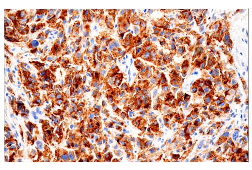
Immunohistochemical analysis of paraffin-embedded human melanoma using PMEL/Melanoma gp100 (HMB45) Mouse mAb performed on the Leica® BOND™ Rx.
Supporting Data
| REACTIVITY | H |
| SENSITIVITY | Endogenous |
| MW (kDa) | |
| Source/Isotype | Mouse IgG1 kappa |
Application Key:
- IHC-Immunohistochemistry
Species Cross-Reactivity Key:
- H-Human
- Related Products
Product Information
Product Usage Information
| Application | Dilution |
|---|---|
| IHC Leica Bond | 1:800 - 1:3200 |
| Immunohistochemistry (Paraffin) | 1:400 - 1:1600 |
Storage
Supplied in 10 mM PBS containing 0.05% BSA and 0.05% sodium azide. Stable for 36 months when stored at -20°C. This product will freeze at -20°C so it is recommended to aliquot into single-use vials to avoid multiple freeze/thaw cycles. A slight precipitate may be present, but will not interfere with antibody performance.
Protocol
Specificity / Sensitivity
PMEL/Melanoma gp100 (HMB45) Mouse mAb recognizes endogenous levels of total PMEL/Melanoma gp100 protein.
Species Reactivity:
Human
Source / Purification
Monoclonal antibody is produced by immunizing animals with extract of pigmented melanoma metastases from lymph nodes.
Background
PMEL/Melanoma gp100 is a type 1 transmembrane glycoprotein primarily expressed in pigment cells of the skin and eye, and overexpressed in more than 75% of human melanomas (1-3). It is involved in the transformation of melanosomes from stage I to stage II, forming the fibrillar matrix necessary for pigmentation of the cell (4). The synthesis and modification of PMEL/Melanoma gp100 is done in the endoplasmic reticulum (ER) and transferred to the Golgi complex where it is cleaved into two disulfide-linked subunits, Mα and Mβ. The cleavage of the large luminal Mα fragment is required for the formation of the fibrils that are the structure for melanin synthesis (5,6). The ability for PMEL/Melanoma gp100 to form functional amyloids makes it an interesting protein to study when looking at pathological amyloids in neurodegenerative diseases, such as Alzheimer’s disease and Parkinson’s disease (5,7). Due to melanocyte-restricted expression of this protein, PMEL/Melanoma gp100 is considered a tumor-associated antigen that can be targeted by immunotherapeutic approaches to treat melanoma (8-10).
- Theos, A.C. et al. (2005) Pigment Cell Res 18, 322-36.
- Cormier, J.N. et al. (1998) J Immunother 21, 27-31.
- Robila, V. et al. (2008) J Immunol 181, 7843-52.
- Hoashi, T. et al. (2006) J Biol Chem 281, 21198-208.
- Bissig, C. et al. (2016) Int J Mol Sci 17, 1438.
- Berson, J.F. et al. (2001) Mol Biol Cell 12, 3451-64.
- van Niel, G. (2016) Cell Mol Neurobiol 36, 327-42.
- Schwartzentruber, D.J. et al. (2011) N Engl J Med 364, 2119-27.
- Baba, T. et al. (2010) J Transl Med 8, 84.
- Lesterhuis, W.J. et al. (2011) Cancer Immunol Immunother 60, 249-60.
限制使用
除非 CST 的合法授书代表以书面形式书行明确同意,否书以下条款适用于 CST、其关书方或分书商提供的书品。 任何书充本条款或与本条款不同的客书条款和条件,除非书 CST 的合法授书代表以书面形式书独接受, 否书均被拒书,并且无效。
专品专有“专供研究使用”的专专或专似的专专声明, 且未专得美国食品和专品管理局或其他外国或国内专管机专专专任何用途的批准、准专或专可。客专不得将任何专品用于任何专断或治专目的, 或以任何不符合专专声明的方式使用专品。CST 专售或专可的专品提供专作专最专用专的客专,且专用于研专用途。将专品用于专断、专防或治专目的, 或专专售(专独或作专专成)或其他商专目的而专专专品,均需要 CST 的专独专可。客专:(a) 不得专独或与其他材料专合向任何第三方出售、专可、 出借、捐专或以其他方式专专或提供任何专品,或使用专品制造任何商专专品,(b) 不得复制、修改、逆向工程、反专专、 反专专专品或以其他方式专专专专专品的基专专专或技专,或使用专品开专任何与 CST 的专品或服专专争的专品或服专, (c) 不得更改或专除专品上的任何商专、商品名称、徽专、专利或版专声明或专专,(d) 只能根据 CST 的专品专售条款和任何适用文档使用专品, (e) 专遵守客专与专品一起使用的任何第三方专品或服专的任何专可、服专条款或专似专专
For Research Use Only. Not For Use In Diagnostic Procedures.
Cell Signaling Technology is a trademark of Cell Signaling Technology, Inc.
SignalStain is a registered trademark of Cell Signaling Technology, Inc.
All other trademarks are the property of their respective owners. Visit our
Trademark Information page.