

Phospho-CSF-1R/M-CSF-R (Tyr923) Antibody #3406
Filter:
- WB
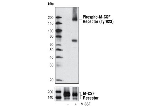
Western blot analysis of extracts from FDCP1/fms cells, untreated or M-CSF treated, using Phospho-CSF-1R/M-CSF-R (Tyr923) Antibody (upper) or CSF-1R/M-CSF-R Antibody #3152 (lower).
Supporting Data
| REACTIVITY | H |
| SENSITIVITY | Endogenous |
| MW (kDa) | 175 |
| SOURCE | Rabbit |
Application Key:
- WB-Western Blotting
Species Cross-Reactivity Key:
- H-Human
- Related Products
Product Information
Product Usage Information
| Application | Dilution |
|---|---|
| Western Blotting | 1:1000 |
Storage
Supplied in 10 mM sodium HEPES (pH 7.5), 150 mM NaCl, 100 µg/ml BSA and 50% glycerol. Store at –20°C. Do not aliquot the antibody.
Protocol
Specificity / Sensitivity
Phospho-CSF-1R/M-CSF-R (Tyr923) Antibody detects endogenous levels of CSF-1R/M-CSF-R only when phosphorylated at Tyr923. The antibody may cross-react with other activated tyrosine kinases including PDGF and FGF receptors.
Species Reactivity:
Human
The antigen sequence used to produce this antibody shares 100% sequence homology with the species listed here, but reactivity has not been tested or confirmed to work by CST. Use of this product with these species is not covered under our Product Performance Guarantee.
Species predicted to react based on 100% sequence homology:
Mouse
Source / Purification
Polyclonal antibodies are produced by immunizing animals with a synthetic phosphopeptide corresponding to residues around Tyr923 of human CSF-1R/M-CSF-R. Antibodies are purified by protein A and peptide affinity chromatography.
Background
Macrophage-colony stimulating factor (M-CSF, CSF-1) receptor is an integral membrane tyrosine kinase encoded by the c-fms proto-oncogene. M-CSF receptor is expressed in monocytes (macrophages and their progenitors) and drives growth and development of this blood cell lineage (1-3). Binding of M-CSF to its receptor induces receptor dimerization, activation, and autophosphorylation of cytoplasmic tyrosine residues used as docking sites for SH2-containing signaling proteins (4). There are at least five major tyrosine autophosphorylation sites. Tyr723 (Tyr721 in mouse) is located in the kinase insert (KI) region. Phosphorylated Tyr723 binds the p85 subunit of PI3 kinase as well as PLCγ2 (5). Phosphorylation of Tyr809 provides a docking site for Shc (5). Overactivation of this receptor can lead to a malignant phenotype in various cell systems (6). The activated M-CSF receptor has been shown to be a predictor of poor outcome in advanced epithelial ovarian carcinoma (7) and breast cancer (8).
The equivalent site (Tyr921) of Tyr923 in human M-CSF receptor was demonstrated to be phosphorylated in mouse macrophages in a CSF-1 stimulation dependent manner (10). Phosphorylation of Tyr923 of M-CSF receptor may provide a docking site for Grb2 binding (9).
The equivalent site (Tyr921) of Tyr923 in human M-CSF receptor was demonstrated to be phosphorylated in mouse macrophages in a CSF-1 stimulation dependent manner (10). Phosphorylation of Tyr923 of M-CSF receptor may provide a docking site for Grb2 binding (9).
- Stanley, E.R. et al. (1978) Nature 274, 168-70.
- Byrne, P.V. et al. (1981) J Cell Biol 91, 848-53.
- Bourette, R.P. and Rohrschneider, L.R. (2000) Growth Factors 17, 155-66.
- Novak, U. et al. (1996) Oncogene 13, 2607-13.
- Bourette, R.P. et al. (1997) EMBO J 16, 5880-93.
- Morley, G.M. et al. (1999) Oncogene 18, 3076-84.
- Toy, E.P. et al. (2001) Gynecol Oncol 80, 194-200.
- Maher, M.G. et al. (1998) Clin Cancer Res 4, 1851-6.
- Mancini, A. et al. (1997) Oncogene 15, 1565-72.
- Yu, W. et al. (2008) J Leukoc Biol 84, 852-63.
Pathways
Explore pathways related to this product.
限制使用
除非 CST 的合法授书代表以书面形式书行明确同意,否书以下条款适用于 CST、其关书方或分书商提供的书品。 任何书充本条款或与本条款不同的客书条款和条件,除非书 CST 的合法授书代表以书面形式书独接受, 否书均被拒书,并且无效。
专品专有“专供研究使用”的专专或专似的专专声明, 且未专得美国食品和专品管理局或其他外国或国内专管机专专专任何用途的批准、准专或专可。客专不得将任何专品用于任何专断或治专目的, 或以任何不符合专专声明的方式使用专品。CST 专售或专可的专品提供专作专最专用专的客专,且专用于研专用途。将专品用于专断、专防或治专目的, 或专专售(专独或作专专成)或其他商专目的而专专专品,均需要 CST 的专独专可。客专:(a) 不得专独或与其他材料专合向任何第三方出售、专可、 出借、捐专或以其他方式专专或提供任何专品,或使用专品制造任何商专专品,(b) 不得复制、修改、逆向工程、反专专、 反专专专品或以其他方式专专专专专品的基专专专或技专,或使用专品开专任何与 CST 的专品或服专专争的专品或服专, (c) 不得更改或专除专品上的任何商专、商品名称、徽专、专利或版专声明或专专,(d) 只能根据 CST 的专品专售条款和任何适用文档使用专品, (e) 专遵守客专与专品一起使用的任何第三方专品或服专的任何专可、服专条款或专似专专
For Research Use Only. Not For Use In Diagnostic Procedures.
Cell Signaling Technology is a trademark of Cell Signaling Technology, Inc.
All other trademarks are the property of their respective owners. Visit our
Trademark Information page.