

NMDAR2B (N59/36.1) Mouse mAb #5580
We recommend the following alternatives
Filter:
- WB
- IP
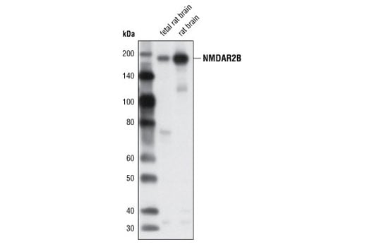
Western blot analysis of extracts from fetal rat brain and rat brain using NMDAR2B (N59/36.1) Mouse mAb.
This product is discontinued
Inquiry Info. # 5580
Please see our recommended alternatives.
Supporting Data
| REACTIVITY | M R |
| SENSITIVITY | Endogenous |
| MW (kDa) | 190 |
| Source/Isotype | Mouse IgG2b |
Application Key:
- WB-Western Blotting
- IP-Immunoprecipitation
Species Cross-Reactivity Key:
- M-Mouse
- R-Rat
Product Information
Product Usage Information
| Application | Dilution |
|---|---|
| Western Blotting | 1:1000 |
| Immunoprecipitation | 1:50 |
Storage
Supplied in 10 mM sodium HEPES (pH 7.5), 150 mM NaCl, 100 µg/ml BSA, 50% glycerol and less than 0.02% sodium azide. Store at –20°C. Do not aliquot the antibody.
Protocol
Specificity / Sensitivity
NMDAR2B (N59/36.1) Mouse mAb detects endogenous levels of total NMDAR2B protein.
Species Reactivity:
Mouse, Rat
Source / Purification
Monoclonal antibody is produced by immunizing animals with a recombinant protein specific to the amino terminus of rat NMDAR2B protein.
Background
N-methyl-D-aspartate receptor (NMDAR) forms a heterodimer of at least one NR1 and one NR2A-D subunit. Multiple receptor isoforms with distinct brain distributions and functional properties arise by selective splicing of the NR1 transcripts and differential expression of the NR2 subunits. NR1 subunits bind the co-agonist glycine and NR2 subunits bind the neurotransmitter glutamate. Activation of the NMDA receptor or opening of the ion channel allows flow of Na+ and Ca2+ ions into the cell, and K+ out of the cell (1). Each subunit has a cytoplasmic domain that can be directly modified by the protein kinase/phosphatase (2). PKC can phosphorylate the NR1 subunit (NMDAR1) of the receptor at Ser890/Ser896, and PKA can phosphorylate NR1 at Ser897 (3). The phosphorylation of NR1 by PKC decreases its affinity for calmodulin, thus preventing the inhibitory effect of calmodulin on NMDAR (4). The phosphorylation of NR1 by PKA probably counteracts the inhibitory effect of calcineurin on the receptor (5). NMDAR mediates long-term potentiation and slow postsynaptic excitation, which play central roles in learning, neurodevelopment, and neuroplasticity (6).
EphrinB2 binding to the receptor EphB leads to the activation of Src family tyrosine kinases, which phosphorylate NMDAR2B at Tyr1252, Tyr1336 and Tyr1472. In turn, phosphorylated NMDAR2B enhances the ability of the functional NMDA receptor to regulate Ca2+ influx in response to glutamate (7).
EphrinB2 binding to the receptor EphB leads to the activation of Src family tyrosine kinases, which phosphorylate NMDAR2B at Tyr1252, Tyr1336 and Tyr1472. In turn, phosphorylated NMDAR2B enhances the ability of the functional NMDA receptor to regulate Ca2+ influx in response to glutamate (7).
- Liu, X.B. et al. (2004) J Neurosci 24, 8885-95.
- Westphal, R.S. et al. (1999) Science 285, 93-6.
- Tingley, W.G. et al. (1997) J Biol Chem 272, 5157-66.
- Hisatsune, C. et al. (1997) J Biol Chem 272, 20805-10.
- Raman, I.M. et al. (1996) Neuron 16, 415-21.
- Makhinson, M. et al. (1999) J Neurosci 19, 2500-10.
- Takasu, M.A. et al. (2002) Science 295, 491-5.
Pathways
Explore pathways related to this product.
限制使用
除非 CST 的合法授书代表以书面形式书行明确同意,否书以下条款适用于 CST、其关书方或分书商提供的书品。 任何书充本条款或与本条款不同的客书条款和条件,除非书 CST 的合法授书代表以书面形式书独接受, 否书均被拒书,并且无效。
专品专有“专供研究使用”的专专或专似的专专声明, 且未专得美国食品和专品管理局或其他外国或国内专管机专专专任何用途的批准、准专或专可。客专不得将任何专品用于任何专断或治专目的, 或以任何不符合专专声明的方式使用专品。CST 专售或专可的专品提供专作专最专用专的客专,且专用于研专用途。将专品用于专断、专防或治专目的, 或专专售(专独或作专专成)或其他商专目的而专专专品,均需要 CST 的专独专可。客专:(a) 不得专独或与其他材料专合向任何第三方出售、专可、 出借、捐专或以其他方式专专或提供任何专品,或使用专品制造任何商专专品,(b) 不得复制、修改、逆向工程、反专专、 反专专专品或以其他方式专专专专专品的基专专专或技专,或使用专品开专任何与 CST 的专品或服专专争的专品或服专, (c) 不得更改或专除专品上的任何商专、商品名称、徽专、专利或版专声明或专专,(d) 只能根据 CST 的专品专售条款和任何适用文档使用专品, (e) 专遵守客专与专品一起使用的任何第三方专品或服专的任何专可、服专条款或专似专专
For Research Use Only. Not For Use In Diagnostic Procedures.
Cell Signaling Technology is a trademark of Cell Signaling Technology, Inc.
All other trademarks are the property of their respective owners. Visit our
Trademark Information page.