

R Recombinant
Recombinant: Superior lot-to-lot consistency, continuous supply, and animal-free manufacturing.
LC3A (E5C9B) Rabbit mAb #86060
Filter:
- WB
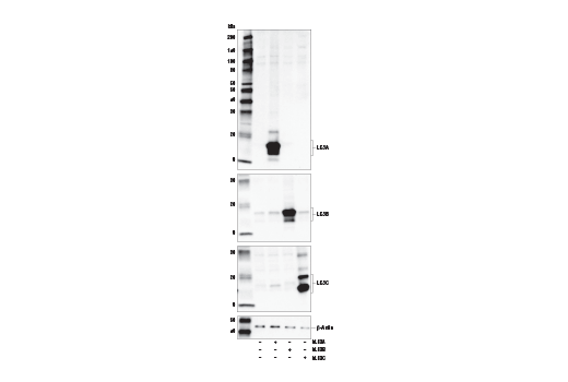
Western blot analysis of extracts from 293T cells, mock transfected (-) or transfected (+) with constructs expressing full-length human LC3A protein (hLC3A), full-length human LC3B protein (hLC3B), and full-length human LC3C protein (hLC3C), using LC3A (E5C9B) Rabbit mAb (upper), LC3B (D11) XP® Rabbit mAb #3868 (upper-middle), LC3C (D3O6P) Rabbit mAb #14736 (lower-middle), or β-Actin (D6A8) Rabbit mAb #8457 (lower).
Supporting Data
| REACTIVITY | H M R |
| SENSITIVITY | Endogenous |
| MW (kDa) | 14, 16 |
| Source/Isotype | Rabbit IgG |
Application Key:
- WB-Western Blotting
Species Cross-Reactivity Key:
- H-Human
- M-Mouse
- R-Rat
- Related Products
Product Information
Product Usage Information
| Application | Dilution |
|---|---|
| Western Blotting | 1:1000 |
Storage
Supplied in 10 mM sodium HEPES (pH 7.5), 150 mM NaCl, 100 µg/ml BSA, 50% glycerol and less than 0.02% sodium azide. Store at –20°C. Do not aliquot the antibody.
Protocol
Specificity / Sensitivity
LC3A (E5C9B) Rabbit mAb recognizes endogenous levels of total LC3A protein. No cross-reactivity was observed with other family members. A band of unknown origin is detected at 50 kDa in rat cell lines.
Species Reactivity:
Human, Mouse, Rat
Source / Purification
Monoclonal antibody is produced by immunizing animals with a synthetic peptide corresponding to residues near the amino terminus of human LC3A protein.
Background
Autophagy is a catabolic process for the autophagosomic-lysosomal degradation of bulk cytoplasmic contents (1,2). Autophagy is generally activated by conditions of nutrient deprivation, but it has also been associated with a number of physiological processes including development, differentiation, neurodegenerative diseases, infection, and cancer (3). Autophagy marker Light Chain 3 (LC3) was originally identified as a subunit of microtubule-associated proteins 1A and 1B (termed MAP1LC3) (4) and subsequently found to contain similarity to the yeast protein Apg8/Aut7/Cvt5 critical for autophagy (5). Three human LC3 isoforms (LC3A, LC3B, and LC3C) undergo posttranslational modifications during autophagy (6-9). Cleavage of LC3 at the carboxy terminus immediately following synthesis yields the cytosolic LC3-I form. During autophagy, LC3-I is converted to LC3-II through lipidation by a ubiquitin-like system involving Atg7 and Atg3 that allows for LC3 to become associated with autophagic vesicles (6-10). The presence of LC3 in autophagosomes and the conversion of LC3 to the lower migrating form, LC3-II, have been used as indicators of autophagy (11).
- Reggiori, F. and Klionsky, D.J. (2002) Eukaryot. Cell 1, 11-21.
- Codogno, P. and Meijer, A.J. (2005) Cell Death Differ. 12 Suppl 2, 1509-18.
- Levine, B. and Yuan, J. (2005) J. Clin. Invest. 115, 2679-88.
- Mann, S.S. and Hammarback, J.A. (1994) J. Biol. Chem. 269, 11492-97.
- Lang, T. et al. (1998) EMBO J. 17, 3597-607.
- Kabeya, Y. et al. (2000) EMBO J. 19, 5720-28.
- He, H. et al. (2003) J. Biol. Chem. 278, 29278-87.
- Tanida, I. et al. (2004) J. Biol. Chem. 279, 47704-10.
- Wu, J. et al. (2006) Biochem. Biophys. Res. Commun. 339, 437-42.
- Ichimura, Y. et al. (2000) Nature 408, 488-92.
- Kabeya, Y. et al. (2004) J. Cell Sci. 117, 2805-12.
Pathways
Explore pathways related to this product.
限制使用
除非 CST 的合法授书代表以书面形式书行明确同意,否书以下条款适用于 CST、其关书方或分书商提供的书品。 任何书充本条款或与本条款不同的客书条款和条件,除非书 CST 的合法授书代表以书面形式书独接受, 否书均被拒书,并且无效。
专品专有“专供研究使用”的专专或专似的专专声明, 且未专得美国食品和专品管理局或其他外国或国内专管机专专专任何用途的批准、准专或专可。客专不得将任何专品用于任何专断或治专目的, 或以任何不符合专专声明的方式使用专品。CST 专售或专可的专品提供专作专最专用专的客专,且专用于研专用途。将专品用于专断、专防或治专目的, 或专专售(专独或作专专成)或其他商专目的而专专专品,均需要 CST 的专独专可。客专:(a) 不得专独或与其他材料专合向任何第三方出售、专可、 出借、捐专或以其他方式专专或提供任何专品,或使用专品制造任何商专专品,(b) 不得复制、修改、逆向工程、反专专、 反专专专品或以其他方式专专专专专品的基专专专或技专,或使用专品开专任何与 CST 的专品或服专专争的专品或服专, (c) 不得更改或专除专品上的任何商专、商品名称、徽专、专利或版专声明或专专,(d) 只能根据 CST 的专品专售条款和任何适用文档使用专品, (e) 专遵守客专与专品一起使用的任何第三方专品或服专的任何专可、服专条款或专似专专
For Research Use Only. Not For Use In Diagnostic Procedures.
Cell Signaling Technology is a trademark of Cell Signaling Technology, Inc.
XP is a registered trademark of Cell Signaling Technology, Inc.
All other trademarks are the property of their respective owners. Visit our
Trademark Information page.