

R Recombinant
Recombinant: Superior lot-to-lot consistency, continuous supply, and animal-free manufacturing.
ROS1 (D4D6®) Rabbit mAb #3287
Filter:
- WB
- IP
- IHC
- IF
- F
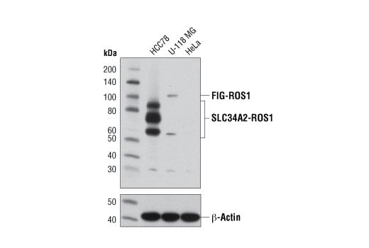
Western blot analysis of extracts from HCC78 (SLC34A2-ROS1), U-118 MG (FIG-ROS1), and HeLa (ROS1 negative) cells using ROS1 (D4D6®) Rabbit mAb (upper) or β-Actin (D6A8) Rabbit mAb #8457 (lower). Note: HCC78 cells express the 85, 70, and 59 kDa forms of the SLC34A2-ROS1 fusion protein (7).
Supporting Data
| REACTIVITY | H |
| SENSITIVITY | Endogenous |
| MW (kDa) | 258, 110, 50-80 |
| Source/Isotype | Rabbit IgG |
Application Key:
- WB-Western Blotting
- IP-Immunoprecipitation
- IHC-Immunohistochemistry
- IF-Immunofluorescence
- F-Flow Cytometry
Species Cross-Reactivity Key:
- H-Human
- Related Products
- Conjugates
Product Information
Product Usage Information
| Application | Dilution |
|---|---|
| Western Blotting | 1:1000 |
| Simple Western™ | 1:10 - 1:50 |
| Immunoprecipitation | 1:50 |
| Immunohistochemistry (Paraffin) | 1:125 - 1:500 |
| Immunofluorescence (Immunocytochemistry) | 1:200 - 1:800 |
| Flow Cytometry (Fixed/Permeabilized) | 1:200 - 1:800 |
Storage
Supplied in 10 mM sodium HEPES (pH 7.5), 150 mM NaCl, 100 µg/ml BSA, 50% glycerol and less than 0.02% sodium azide. Store at –20°C. Do not aliquot the antibody.
Protocol
Specificity / Sensitivity
ROS1 (D4D6®) Rabbit mAb recognizes endogenous levels of total ROS1 protein. This antibody does not cross-react with other related proteins when analyzed by western blot. Please note that staining may be observed in ROS1 rearranged lung carcinomas, macrophages/giant cells, reactive type II pneumocyte hyperplasia, and the epithelium in areas of bronchiolar metaplasia. Staining of unknown specificity has been observed in cholangiocarcinoma, hepatocellular carcinoma, and kidney tissues.
Species Reactivity:
Human
Source / Purification
Monoclonal antibody is produced by immunizing animals with a protein corresponding to residues in the carboxy terminal domain of the human ROS1 protein.
Background
ROS1, an orphan receptor tyrosine kinase of the insulin receptor family, was initially identified as a homolog of v-ros from the UR2 sarcoma virus (1). ROS1 consists of a large extracellular domain that is composed of six fibronectin repeats, a transmembrane domain, and a C-terminal kinase domain. Being an orphan receptor, the functions of ROS1 are not well known, though it has been shown to play an important role in differentiation of epididymal epithelium (2). The first oncogenic fusion of ROS1, FIG-ROS1, was initially identified by research studies in glioblastoma (3), and subsequent studies have found this fusion in cholangiocarcinoma (4), ovarian cancer (5), and non-small cell lung cancer (NSCLC) (6). Investigators have found additional oncogenic ROS1 fusion proteins in NSCLC (at a frequency of ~1.6%), where the ROS1 kinase domain is fused to the amino-terminal region of several different proteins, including CD74 and SLC34A2 (6-8). ROS1 fusion proteins activate the SHP-2 phosphatase, PI3K/Akt/mTOR, Erk, and Stat3 pathways (3,4,9). There are two autophosphorylation sites (Tyr2274, Tyr2334) downstream of the kinase domain of ROS1, either of which may serve as biomarkers of ROS1 kinase activity, including that of ROS1 fusion proteins (10).
- Matsushime, H. et al. (1986) Mol Cell Biol 6, 3000-4.
- Yeung, C.H. et al. (1999) Biol Reprod 61, 1062-9.
- Charest, A. et al. (2003) Genes Chromosomes Cancer 37, 58-71.
- Gu, T.L. et al. (2011) PLoS One 6, e15640.
- Birch, A.H. et al. (2011) PLoS One 6, e28250.
- Rimkunas, V.M. et al. (2012) Clin Cancer Res 18, 4449-57.
- Rikova, K. et al. (2007) Cell 131, 1190-203.
- Stumpfova, M. and Jänne, P.A. (2012) Clin Cancer Res 18, 4222-4.
- Jun, H.J. et al. (2012) Cancer Res 72, 3764-74.
- Zou, H.Y. et al. (2015) Proc Natl Acad Sci U S A 112, 3493-8.
Pathways
Explore pathways related to this product.
限制使用
除非 CST 的合法授书代表以书面形式书行明确同意,否书以下条款适用于 CST、其关书方或分书商提供的书品。 任何书充本条款或与本条款不同的客书条款和条件,除非书 CST 的合法授书代表以书面形式书独接受, 否书均被拒书,并且无效。
专品专有“专供研究使用”的专专或专似的专专声明, 且未专得美国食品和专品管理局或其他外国或国内专管机专专专任何用途的批准、准专或专可。客专不得将任何专品用于任何专断或治专目的, 或以任何不符合专专声明的方式使用专品。CST 专售或专可的专品提供专作专最专用专的客专,且专用于研专用途。将专品用于专断、专防或治专目的, 或专专售(专独或作专专成)或其他商专目的而专专专品,均需要 CST 的专独专可。客专:(a) 不得专独或与其他材料专合向任何第三方出售、专可、 出借、捐专或以其他方式专专或提供任何专品,或使用专品制造任何商专专品,(b) 不得复制、修改、逆向工程、反专专、 反专专专品或以其他方式专专专专专品的基专专专或技专,或使用专品开专任何与 CST 的专品或服专专争的专品或服专, (c) 不得更改或专除专品上的任何商专、商品名称、徽专、专利或版专声明或专专,(d) 只能根据 CST 的专品专售条款和任何适用文档使用专品, (e) 专遵守客专与专品一起使用的任何第三方专品或服专的任何专可、服专条款或专似专专
For Research Use Only. Not For Use In Diagnostic Procedures.
Cell Signaling Technology is a trademark of Cell Signaling Technology, Inc.
Alexa Fluor is a registered trademark of Life Technologies Corporation.
All other trademarks are the property of their respective owners. Visit our
Trademark Information page.