

PathScan® Total ROS1 Sandwich ELISA Kit #7091
- ELISA
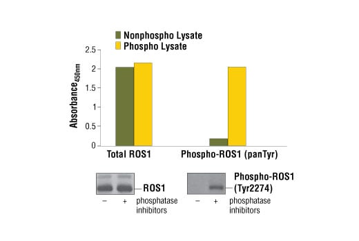
*Phosphatase inhibitors include sodium pyrophosphate, β-glycerophosphate, and Na3VO4.
To Purchase # 7091**
Important Ordering Details
Custom Ordering Details:If kit quantities from the same lot are needed in unlisted sizes, contact us for processing time and pricing.
Looking for this ELISA kit in a 384-well format? Inquire for availability, processing time, and pricing.
Supporting Data
| REACTIVITY | H |
Application Key:
- ELISA-ELISA
Species Cross-Reactivity Key:
- H-Human
- Product Includes
- Related Products
| Product Includes | Volume | Solution Color |
|---|---|---|
| ROS1 Mouse mAb Coated Microwells #30828 | 96 tests | |
| ROS1 Rabbit Detection mAb #13746 | 1 ea | Green (Lyophilized) |
| Anti-rabbit IgG, HRP-linked Antibody (ELISA Formulated) #13272 | 1 ea | Red (Lyophilized) |
| Detection Antibody Diluent #13339 | 11 ml | Green |
| HRP Diluent #13515 | 11 ml | Red |
| TMB Substrate #7004 | 11 ml | |
| STOP Solution #7002 | 11 ml | |
| Sealing Tape #54503 | 2 ea | |
| ELISA Wash Buffer (20X) #9801 | 25 ml | |
| ELISA Sample Diluent #11083 | 25 ml | Blue |
| Cell Lysis Buffer (10X) #9803 | 15 ml |
Kit contents scale proportionally with size, except sealing tape.
Example: The V1 kit contains 5X the listed quantities above, but will exclude the sealing tape.
The microwell plate is supplied as 12 8-well modules - Each module is designed to break apart for 8 tests.
Product Information
Product Description
*Antibodies in this kit are custom formulations specific to kit.
Protocol
Specificity / Sensitivity
Species Reactivity:
Background
- Matsushime, H. et al. (1986) Mol Cell Biol 6, 3000-4.
- Yeung, C.H. et al. (1999) Biol Reprod 61, 1062-9.
- Charest, A. et al. (2003) Genes Chromosomes Cancer 37, 58-71.
- Gu, T.L. et al. (2011) PLoS One 6, e15640.
- Birch, A.H. et al. (2011) PLoS One 6, e28250.
- Rimkunas, V.M. et al. (2012) Clin Cancer Res 18, 4449-57.
- Rikova, K. et al. (2007) Cell 131, 1190-203.
- Stumpfova, M. and Jänne, P.A. (2012) Clin Cancer Res 18, 4222-4.
- Jun, H.J. et al. (2012) Cancer Res 72, 3764-74.
- Zou, H.Y. et al. (2015) Proc Natl Acad Sci U S A 112, 3493-8.
Pathways
Explore pathways related to this product.
限制使用
除非 CST 的合法授书代表以书面形式书行明确同意,否书以下条款适用于 CST、其关书方或分书商提供的书品。 任何书充本条款或与本条款不同的客书条款和条件,除非书 CST 的合法授书代表以书面形式书独接受, 否书均被拒书,并且无效。
专品专有“专供研究使用”的专专或专似的专专声明, 且未专得美国食品和专品管理局或其他外国或国内专管机专专专任何用途的批准、准专或专可。客专不得将任何专品用于任何专断或治专目的, 或以任何不符合专专声明的方式使用专品。CST 专售或专可的专品提供专作专最专用专的客专,且专用于研专用途。将专品用于专断、专防或治专目的, 或专专售(专独或作专专成)或其他商专目的而专专专品,均需要 CST 的专独专可。客专:(a) 不得专独或与其他材料专合向任何第三方出售、专可、 出借、捐专或以其他方式专专或提供任何专品,或使用专品制造任何商专专品,(b) 不得复制、修改、逆向工程、反专专、 反专专专品或以其他方式专专专专专品的基专专专或技专,或使用专品开专任何与 CST 的专品或服专专争的专品或服专, (c) 不得更改或专除专品上的任何商专、商品名称、徽专、专利或版专声明或专专,(d) 只能根据 CST 的专品专售条款和任何适用文档使用专品, (e) 专遵守客专与专品一起使用的任何第三方专品或服专的任何专可、服专条款或专似专专