

R Recombinant
Recombinant: Superior lot-to-lot consistency, continuous supply, and animal-free manufacturing.
Pan-Keratin (C11) Mouse mAb (Alexa Fluor® 594 Conjugate) #31842
Filter:
- IF
- F
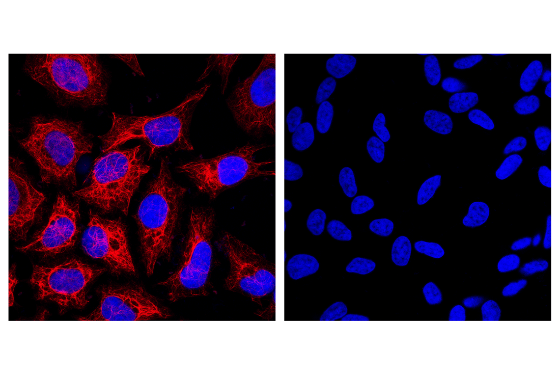
Confocal immunofluorescent analysis of HeLa cells (left, positive) and SH-SY5Y cells (right, negative) using Pan-Keratin (C11) Mouse mAb (Alexa Fluor® 594 Conjugate) (red). Nuclei were labeled with DAPI #4083 (blue).
Supporting Data
| REACTIVITY | H M R Mk |
| SENSITIVITY | Endogenous |
| MW (kDa) | |
| Source/Isotype | Mouse IgG1 |
Application Key:
- IF-Immunofluorescence
- F-Flow Cytometry
Species Cross-Reactivity Key:
- H-Human
- M-Mouse
- R-Rat
- Mk-Monkey
- Related Products
Product Information
Product Description
This Cell Signaling Technology antibody is conjugated to Alexa Fluor® 594 fluorescent dye and tested in-house for direct flow cytometric and immunofluorescent analysis in human cells. This antibody is expected to exhibit the same species cross-reactivity as the unconjugated Pan-Keratin (C11) Mouse mAb #4545.
Product Usage Information
| Application | Dilution |
|---|---|
| Immunofluorescence (Immunocytochemistry) | 1:50 - 1:200 |
| Flow Cytometry (Fixed/Permeabilized) | 1:50 |
Storage
Supplied in PBS (pH 7.2), less than 0.1% sodium azide and 2 mg/ml BSA. Store at 4°C. Do not aliquot the antibody. Protect from light. Do not freeze.
Protocol
Specificity / Sensitivity
Pan-Keratin (C11) Mouse mAb (Alexa Fluor® 594 Conjugate) detects endogenous levels of total keratin 4, 5, 6, 8, 10, 13, and 18. The antibody does not cross-react with other keratins.
Species Reactivity:
Human, Mouse, Rat, Monkey
Source / Purification
Monoclonal antibody (isotype: IgG1) is produced by immunizing a BALB/c mouse with a cytoskeleton preparation from A-431 cells.
Background
Keratins (cytokeratins) are intermediate filament proteins that are mainly expressed in epithelial cells. Keratin heterodimers composed of an acidic keratin (or type I keratin, keratins K9-K28) and a basic keratin (or type II keratin, keratins K1-K8 and K71-K80) assemble to form filaments. Keratin isoforms demonstrate tissue- and differentiation-specific profiles that make them useful as research and clinical biomarkers (1,2).
Dysregulation/mutations in keratin genes can lead to a variety of disorders affecting the skin, hair, nails, and other epithelial tissues (3). While expression of keratins can be variable, immunohistochemical staining of keratins is widely used to help in the identification and classification of epithelial tumors, and may also provide prognostic information.
Keratins 8 and 18 (K8/K18) are expressed in simple epithelia of normal tissue, as well as in adenocarcinomas of the breast, lung, ovary, and gastrointestinal tract. Keratin 17 is expressed in basal keratinocytes of stratified epithelia, hair follicles, and sebaceous glands. Onset of keratin 17 expression coincides with the definition of major epithelial lineages during skin development (4). Keratin 14 (K14) is expressed in basal cells of stratified epithelia, and in basal-like subtypes of breast cancer and squamous cell carcinomas. Keratin 19 (K19) is expressed in glandular epithelia, including the liver, gallbladder, and pancreas, as well as in adenocarcinomas of the breast, thyroid, and bile duct. Keratin 20 (K20) is expressed in gastrointestinal epithelium, urothelium, and Merkel cells in the skin, as well as in colorectal carcinomas and some urothelial carcinomas. Keratin 5/6 (K5/6) is expressed in basal cells of stratified epithelia, including the skin, prostate, and breast, as well as in basal-like breast cancers, squamous cell carcinomas, and some lung carcinomas. Keratin 7 (K7) is expressed in glandular epithelia, such as those in the lung, breast, and female reproductive tract, as well as in adenocarcinomas of the lung, breast, and ovary (5,6).
Keratins, particularly K8, K18, and K19, serve as biomarkers for identification of circulating tumor cells (CTCs) (5).
Post-translational modifications, including phosphorylation, acetylation, ubiquitylation, sumoylation, glycosylation, and transamidation, have been shown to affect the functions of keratins in normal and disease states (6). Understanding the molecular mechanisms underlying these PTMs may provide insights into cancer pathogenesis.
- Chang, L. and Goldman, R.D. (2004) Nat Rev Mol Cell Biol 5, 601-13.
- Schweizer, J. et al. (2006) J Cell Biol 174, 169-74.
- Sarma, A. (2022) Int J Biol Macromol 219, 395-413.
- McGowan, K.M. and Coulombe, P.A. (1998) J Cell Biol 143, 469-86.
- Werner, S. et al. (2020) Mol Aspects Med 72, 100817.
- Dmello, C. et al. (2019) J Biosci 44, 33.
限制使用
除非 CST 的合法授书代表以书面形式书行明确同意,否书以下条款适用于 CST、其关书方或分书商提供的书品。 任何书充本条款或与本条款不同的客书条款和条件,除非书 CST 的合法授书代表以书面形式书独接受, 否书均被拒书,并且无效。
专品专有“专供研究使用”的专专或专似的专专声明, 且未专得美国食品和专品管理局或其他外国或国内专管机专专专任何用途的批准、准专或专可。客专不得将任何专品用于任何专断或治专目的, 或以任何不符合专专声明的方式使用专品。CST 专售或专可的专品提供专作专最专用专的客专,且专用于研专用途。将专品用于专断、专防或治专目的, 或专专售(专独或作专专成)或其他商专目的而专专专品,均需要 CST 的专独专可。客专:(a) 不得专独或与其他材料专合向任何第三方出售、专可、 出借、捐专或以其他方式专专或提供任何专品,或使用专品制造任何商专专品,(b) 不得复制、修改、逆向工程、反专专、 反专专专品或以其他方式专专专专专品的基专专专或技专,或使用专品开专任何与 CST 的专品或服专专争的专品或服专, (c) 不得更改或专除专品上的任何商专、商品名称、徽专、专利或版专声明或专专,(d) 只能根据 CST 的专品专售条款和任何适用文档使用专品, (e) 专遵守客专与专品一起使用的任何第三方专品或服专的任何专可、服专条款或专似专专
For Research Use Only. Not For Use In Diagnostic Procedures.
Cell Signaling Technology is a trademark of Cell Signaling Technology, Inc.
This product is provided under an intellectual property license from Life Technologies Corporation. The transfer of this product is conditioned on the buyer using the purchased product solely in research conducted by the buyer, excluding contract research or any fee for service research, and the buyer must not (1) use this product or its components for (a) diagnostic, therapeutic or prophylactic purposes; (b) testing, analysis or screening services, or information in return for compensation on a per-test basis; or (c) manufacturing or quality assurance or quality control, and/or (2) sell or transfer this product or its components for resale, whether or not resold for use in research. For information on purchasing a license to this product for purposes other than as described above, contact Life Technologies Corporation, 5791 Van Allen Way, Carlsbad, CA 92008 USA or outlicensing@lifetech.com.
All other trademarks are the property of their respective owners. Visit our
Trademark Information page.