

Phospho-RelB (Ser552) Antibody #4999
Filter:
- WB
- IP
- IF
- F
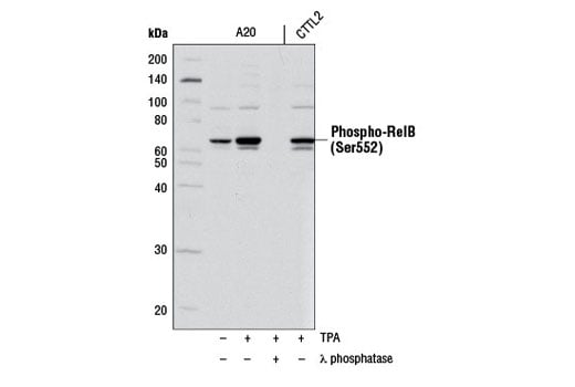
Western blot analysis of extracts from A20 and CTTL2 cells, untreated or treated with TPA #4174 (200nM, 30 minutes) alone or with λ phosphatase, using Phospho-RelB (Ser552) Antibody.
Supporting Data
| REACTIVITY | H M |
| SENSITIVITY | Endogenous |
| MW (kDa) | 70 |
| SOURCE | Rabbit |
Application Key:
- WB-Western Blotting
- IP-Immunoprecipitation
- IF-Immunofluorescence
- F-Flow Cytometry
Species Cross-Reactivity Key:
- H-Human
- M-Mouse
- Related Products
Product Information
Product Usage Information
| Application | Dilution |
|---|---|
| Western Blotting | 1:1000 |
| Immunoprecipitation | 1:100 |
| Immunofluorescence (Immunocytochemistry) | 1:100 |
| Flow Cytometry (Fixed/Permeabilized) | 1:400 |
Storage
Supplied in 10 mM sodium HEPES (pH 7.5), 150 mM NaCl, 100 µg/ml BSA and 50% glycerol. Store at –20°C. Do not aliquot the antibody.
Protocol
Specificity / Sensitivity
Phospho-RelB (Ser552) Antibody detects endogenous levels of RelB only when phosphorylated at Ser552.
Species Reactivity:
Human, Mouse
The antigen sequence used to produce this antibody shares 100% sequence homology with the species listed here, but reactivity has not been tested or confirmed to work by CST. Use of this product with these species is not covered under our Product Performance Guarantee.
Species predicted to react based on 100% sequence homology:
Rat, Monkey, Bovine, Dog
Source / Purification
Polyclonal antibodies are produced by immunizing animals with a synthetic phosphopeptide corresponding to residues surrounding Ser552 of mouse RelB. Antibodies are purified by protein A and peptide affinity chromatography.
Background
Transcription factors of the nuclear factor κB (NF-κB)/Rel family play a pivotal role in inflammatory and immune responses (1,2). There are five family members in mammals: RelA, c-Rel, RelB, NF-κB1 (p105/p50), and NF-κB2 (p100/p52). Both p105 and p100 are proteolytically processed by the proteasome to produce p50 and p52, respectively. Rel proteins bind p50 and p52 to form dimeric complexes that bind DNA and regulate transcription. In unstimulated cells, NF-κB is sequestered in the cytoplasm by IκB inhibitory proteins (3-5). NF-κB-activating agents can induce the phosphorylation of IκB proteins, targeting them for rapid degradation through the ubiquitin-proteasome pathway and releasing NF-κB to enter the nucleus where it regulates gene expression (6-8). NIK and IKKα (IKK1) regulate the phosphorylation and processing of NF-κB2 (p100) to produce p52, which translocates to the nucleus (9-11).
RelB, which is generally activated by non-canonical signaling, forms heterodimers with either p50 or p52 NF-κB subunits to regulate transcription (12,13). RelB null mice are significantly impaired in inflammatory responses and hematopoietic differentiation (14,15). Phosphorlyation at Thr84 and Ser552 results in proteosomal degradation (16).
RelB, which is generally activated by non-canonical signaling, forms heterodimers with either p50 or p52 NF-κB subunits to regulate transcription (12,13). RelB null mice are significantly impaired in inflammatory responses and hematopoietic differentiation (14,15). Phosphorlyation at Thr84 and Ser552 results in proteosomal degradation (16).
- Baeuerle, P.A. and Henkel, T. (1994) Annu Rev Immunol 12, 141-79.
- Baeuerle, P.A. and Baltimore, D. (1996) Cell 87, 13-20.
- Haskill, S. et al. (1991) Cell 65, 1281-9.
- Thompson, J.E. et al. (1995) Cell 80, 573-82.
- Whiteside, S.T. et al. (1997) EMBO J 16, 1413-26.
- Traenckner, E.B. et al. (1995) EMBO J 14, 2876-83.
- Scherer, D.C. et al. (1995) Proc Natl Acad Sci USA 92, 11259-63.
- Chen, Z.J. et al. (1996) Cell 84, 853-62.
- Senftleben, U. et al. (2001) Science 293, 1495-9.
- Coope, H.J. et al. (2002) EMBO J 21, 5375-85.
- Xiao, G. et al. (2001) Mol Cell 7, 401-9.
- Ryseck, R.P. et al. (1992) Mol Cell Biol 12, 674-84.
- Bours, V. et al. (1994) Oncogene 9, 1699-702.
- Weih, F. et al. (1995) Cell 80, 331-40.
- Burkly, L. et al. (1995) Nature 373, 531-6.
- Marienfeld, R. et al. (2001) Oncogene 20, 8142-7.
Pathways
Explore pathways related to this product.
限制使用
除非 CST 的合法授书代表以书面形式书行明确同意,否书以下条款适用于 CST、其关书方或分书商提供的书品。 任何书充本条款或与本条款不同的客书条款和条件,除非书 CST 的合法授书代表以书面形式书独接受, 否书均被拒书,并且无效。
专品专有“专供研究使用”的专专或专似的专专声明, 且未专得美国食品和专品管理局或其他外国或国内专管机专专专任何用途的批准、准专或专可。客专不得将任何专品用于任何专断或治专目的, 或以任何不符合专专声明的方式使用专品。CST 专售或专可的专品提供专作专最专用专的客专,且专用于研专用途。将专品用于专断、专防或治专目的, 或专专售(专独或作专专成)或其他商专目的而专专专品,均需要 CST 的专独专可。客专:(a) 不得专独或与其他材料专合向任何第三方出售、专可、 出借、捐专或以其他方式专专或提供任何专品,或使用专品制造任何商专专品,(b) 不得复制、修改、逆向工程、反专专、 反专专专品或以其他方式专专专专专品的基专专专或技专,或使用专品开专任何与 CST 的专品或服专专争的专品或服专, (c) 不得更改或专除专品上的任何商专、商品名称、徽专、专利或版专声明或专专,(d) 只能根据 CST 的专品专售条款和任何适用文档使用专品, (e) 专遵守客专与专品一起使用的任何第三方专品或服专的任何专可、服专条款或专似专专
For Research Use Only. Not For Use In Diagnostic Procedures.
Cell Signaling Technology is a trademark of Cell Signaling Technology, Inc.
All other trademarks are the property of their respective owners. Visit our
Trademark Information page.