Revision 3

#46008

Store at -20C

CST Logo
Orders:

877-616-CELL (2355)

[email protected]

Support:

877-678-TECH (8324)

3 Trask Lane | Danvers | Massachusetts | 01923 | USA

For Research Use Only. Not for Use in Diagnostic Procedures.

Applications:
W, IP, IHC-P, IF-F, IF-IC, FC-FP

Reactivity:
H M R

Sensitivity:
Endogenous

MW (kDa):
62-82

Source/Isotype:
Rabbit IgG

UniProt ID:
#P15884

Entrez-Gene Id:
6925

Product Usage Information

Application Dilution
Western Blotting 1:1000
Immunoprecipitation 1:50
Immunohistochemistry (Paraffin) 1:1500 - 1:6000
Immunofluorescence (Frozen) 1:50 - 1:200
Immunofluorescence (Immunocytochemistry) 1:1600
Flow Cytometry (Fixed/Permeabilized) 1:3200 - 1:6400

Storage

Supplied in 10 mM sodium HEPES (pH 7.5), 150 mM NaCl, 100 µg/mL BSA, 50% glycerol, and less than 0.02% sodium azide. Store at –20°C. Do not aliquot the antibody.

Specificity/Sensitivity

TCF4/ITF2 (E3X2Y) Rabbit mAb recognizes endogenous levels of total TCF4/ITF2 protein. This antibody is not recommended for immunohistochemical analysis of human tissues. TCF4/ITF2 (E3X2Y) Rabbit mAb is predicted to detect all 16 isoforms of human TCF4/ITF2 protein produced by alternative splicing described on Uniprot (P15884-1 through P15884-16).

Species predicted to react based on 100% sequence homology

Bovine, Dog, Pig, Horse, Rabbit

Source / Purification

Monoclonal antibody is produced by immunizing animals with a synthetic peptide corresponding to residues surrounding Pro653 of human TCF4/ITF2 protein.

Background

Transcription factor 4 (TCF4), also known as immunoglobulin transcription factor 2 (ITF2), is a member of the basic helix-loop-helix family and interacts with Ephrussi-box (E-box) binding sites (1,2). TCF4 is broadly expressed, and due to alternative splicing, transcripts can give rise to over a dozen different isoforms (3). It plays a key role in neuronal development and has been implicated in a variety of developmental disorders, including Pitt-Hopkins syndrome, schizophrenia, and autism (4-9). In addition to its functions in the brain, TCF4 is essential for the development of plasmacytoid dendritic cells (10-12). Finally, in the context of cancer, TCF4 expression may be associated with divergent patient outcomes; higher TCF4 expression is associated with increased survival in non-small cell lung cancer, yet the protein has also recently been reported to confer immunotherapy resistance in melanoma (13,14).

Species Reactivity

Species reactivity is determined by testing in at least one approved application (e.g., western blot).

Western Blot Buffer

IMPORTANT: For western blots, incubate membrane with diluted primary antibody in 5% w/v BSA, 1X TBS, 0.1% Tween® 20 at 4°C with gentle shaking, overnight.

Applications Key

W: Western Blotting IP: Immunoprecipitation IHC-P: Immunohistochemistry (Paraffin) IF-F: Immunofluorescence (Frozen) FC-FP: Flow Cytometry (Fixed/Permeabilized)

Cross-Reactivity Key

H: Human M: Mouse R: Rat Hm: Hamster Mk: Monkey Vir: Virus Mi: Mink C: Chicken Dm: D. melanogaster X: Xenopus Z: Zebrafish B: Bovine Dg: Dog Pg: Pig Sc: S. cerevisiae Ce: C. elegans Hr: Horse GP: Guinea Pig Rab: Rabbit G: Goat All: All Species Expected

Trademarks and Patents

Cell Signaling Technology is a trademark of Cell Signaling Technology, Inc.

All other trademarks are the property of their respective owners. Visit cellsignal.com/trademarks for more information.

限制使用

除非 CST 的合法授书代表以书面形式书行明确同意,否书以下条款适用于 CST、其关书方或分书商提供的书品。 任何书充本条款或与本条款不同的客书条款和条件,除非书 CST 的合法授书代表以书面形式书独接受, 否书均被拒书,并且无效。

专品专有“专供研究使用”的专专或专似的专专声明, 且未专得美国食品和专品管理局或其他外国或国内专管机专专专任何用途的批准、准专或专可。客专不得将任何专品用于任何专断或治专目的, 或以任何不符合专专声明的方式使用专品。CST 专售或专可的专品提供专作专最专用专的客专,且专用于研专用途。将专品用于专断、专防或治专目的, 或专专售(专独或作专专成)或其他商专目的而专专专品,均需要 CST 的专独专可。客专:(a) 不得专独或与其他材料专合向任何第三方出售、专可、 出借、捐专或以其他方式专专或提供任何专品,或使用专品制造任何商专专品,(b) 不得复制、修改、逆向工程、反专专、 反专专专品或以其他方式专专专专专品的基专专专或技专,或使用专品开专任何与 CST 的专品或服专专争的专品或服专, (c) 不得更改或专除专品上的任何商专、商品名称、徽专、专利或版专声明或专专,(d) 只能根据 CST 的专品专售条款和任何适用文档使用专品 , (e) 专遵守客专与专品一起使用的任何第三方专品或服专的任何专可、服专条款或专似专专

Orders: 877-616-CELL (2355) [email protected] Support: 877-678-TECH (8324) [email protected] Web: cellsignal.com
For Research Use Only. Not for Use in Diagnostic Procedures.

Revision 3

CST Logo
Western blot analysis of extracts from various human cell lines using TCF4/ITF2 (E3X2Y) Rabbit mAb (upper) or GAPDH (D16H11) XP® Rabbit mAb #5174 (lower). Negative expression of TCF4/ITF2 protein in Kasumi-1, A549, and JAR cells is consistent with the predicted expression pattern.
Western Blotting Image 1: TCF4/ITF2 (E3X2Y) Rabbit mAb
Western blot analysis of extracts from various human, mouse, and rat cell lines and mouse and rat brain tissues using TCF4/ITF2 (E3X2Y) Rabbit mAb (upper) or GAPDH (D16H11) XP® Rabbit mAb #5174 (lower). Negative expression of TCF4/ITF2 protein in Kasumi-1 and EL4 cells is consistent with the predicted expression pattern.
Western Blotting Image 2: TCF4/ITF2 (E3X2Y) Rabbit mAb
Western blot analysis of extracts from 293T cells, mock transfected (-) or transfected with a construct expressing Myc/DDK-tagged full-length human TCF4/ITF2 protein (hTCF4/ITF2-Myc/DDK; +), using TCF4/ITF2 (E3X2Y) Rabbit mAb (upper) or GAPDH (D16H11) XP® Rabbit mAb #5174 (lower).
Western Blotting Image 3: TCF4/ITF2 (E3X2Y) Rabbit mAb
Orders: 877-616-CELL (2355) [email protected] Support: 877-678-TECH (8324) [email protected] Web: cellsignal.com
For Research Use Only. Not for Use in Diagnostic Procedures.

Revision 3

CST Logo
Immunoprecipitation of TCF4/ITF2 protein from KMS-11 cell extracts. Lane 1 is 10% input, lane 2 is Rabbit (DA1E) mAb IgG XP® Isotope Control #3900, and lane 3 is TCF4/ITF2 (E3X2Y) Rabbit mAb. Western blot analysis was performed using TCF4/ITF2 (E3X2Y) Rabbit mAb. Mouse Anti-Rabbit IgG (Light-Chain Specific) (D4W3E) mAb (HRP Conjugate) #93702 was used as a secondary antibody.
Immunoprecipitation Image 1: TCF4/ITF2 (E3X2Y) Rabbit mAb
Immunohistochemical analysis of paraffin-embedded LL/2 syngeneic tumor using TCF4/ITF2 (E3X2Y) Rabbit mAb.
Immunohistochemistry Image 1: TCF4/ITF2 (E3X2Y) Rabbit mAb

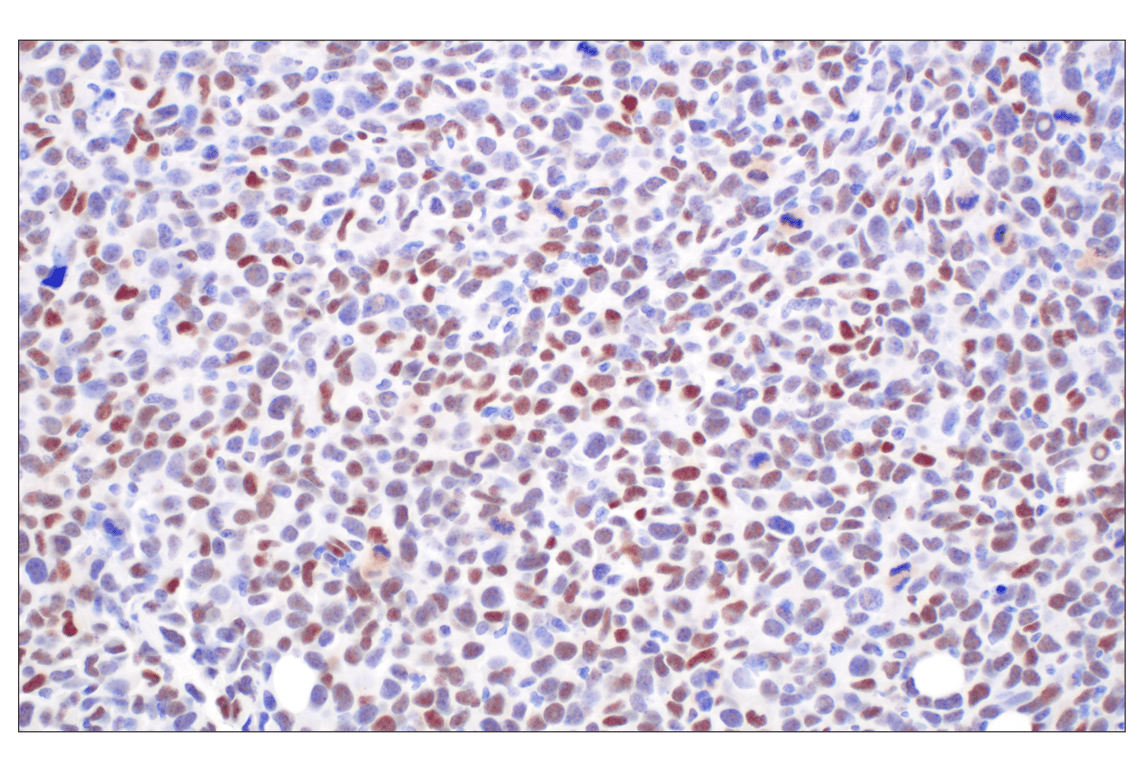
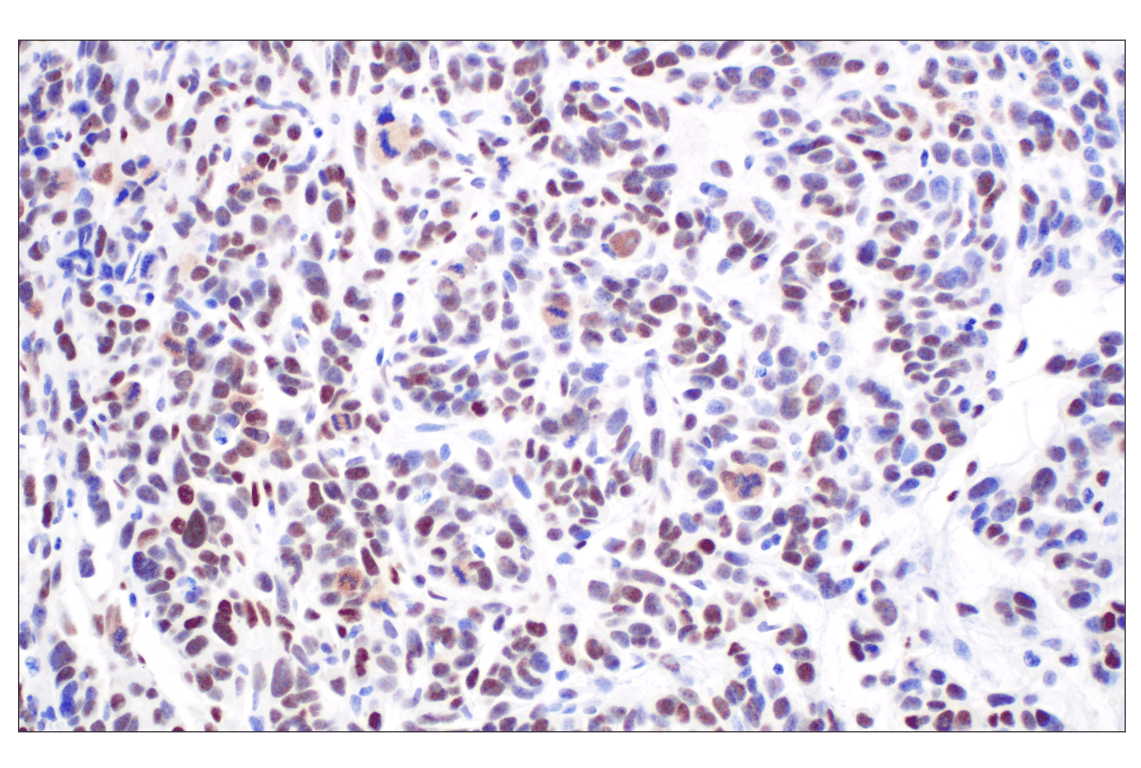
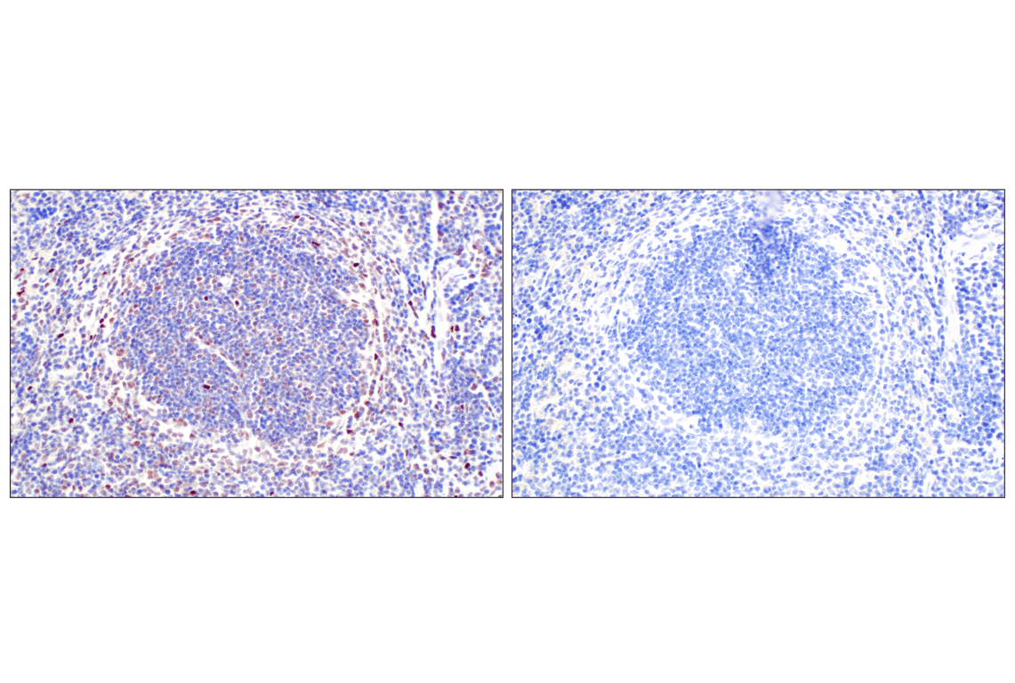
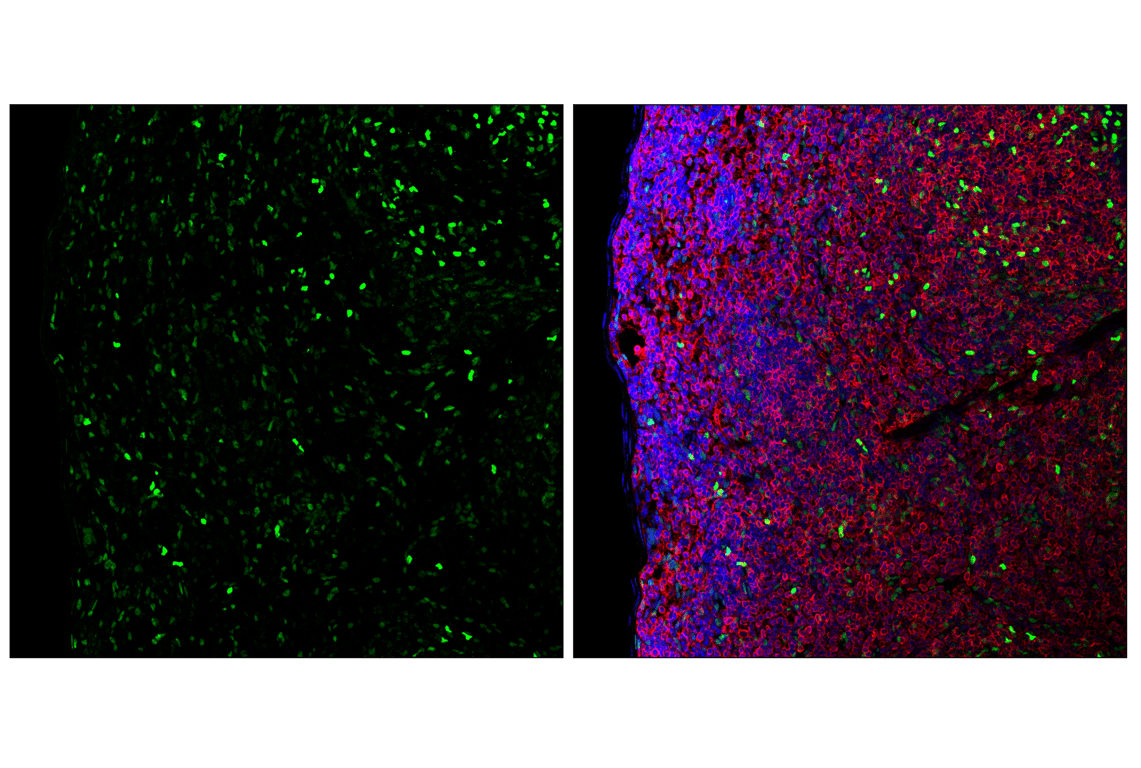
Immunohistochemical analysis of paraffin-embedded A20 syngeneic tumor using TCF4/ITF2 (E3X2Y) Rabbit mAb.

Immunohistochemistry Image 2: TCF4/ITF2 (E3X2Y) Rabbit mAb
Orders: 877-616-CELL (2355) [email protected] Support: 877-678-TECH (8324) [email protected] Web: cellsignal.com
For Research Use Only. Not for Use in Diagnostic Procedures.

Revision 3

CST Logo

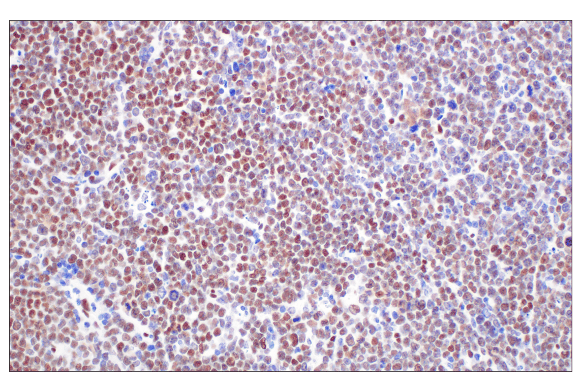
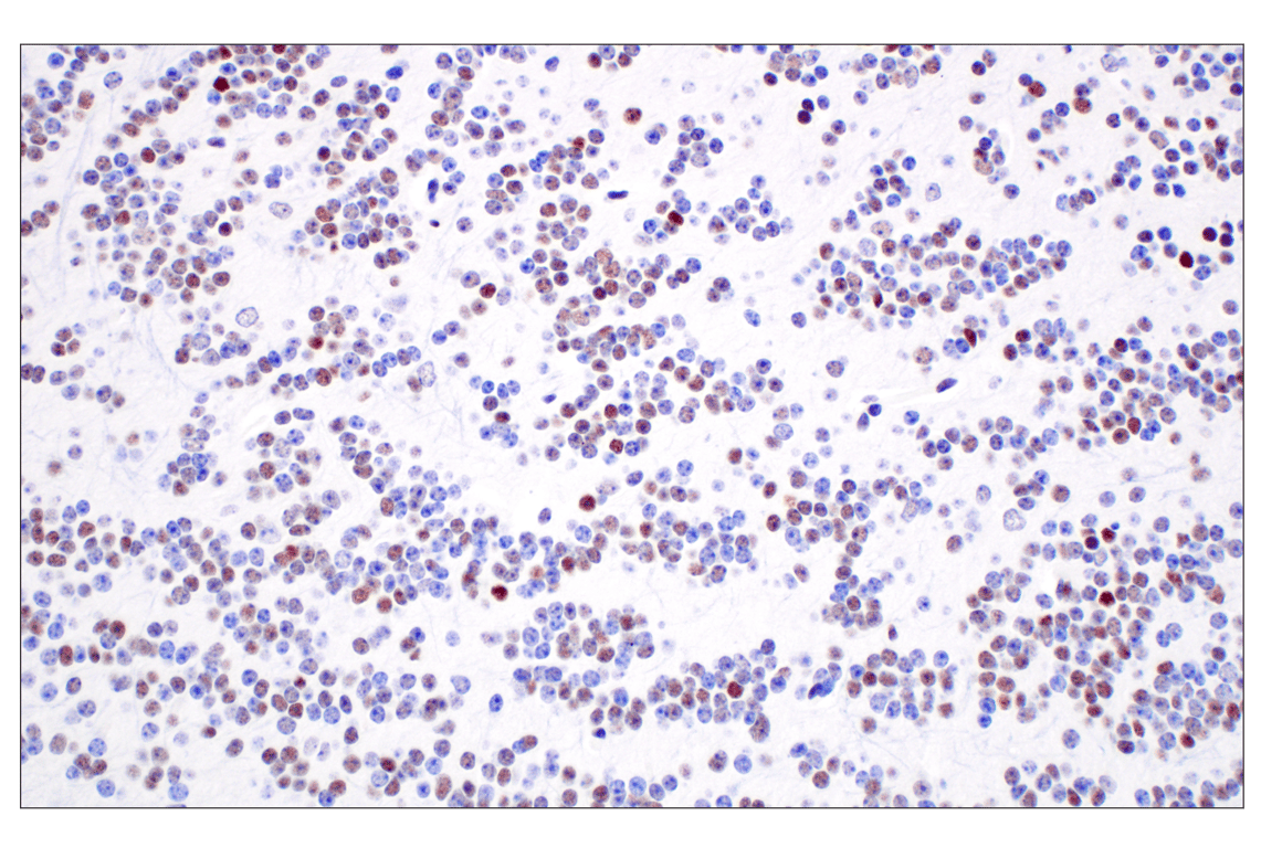
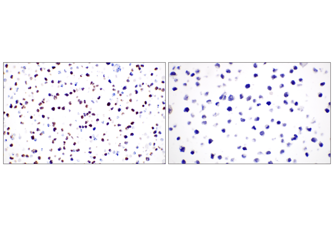
Immunohistochemical analysis of paraffin-embedded GL261 syngeneic tumor using TCF4/ITF2 (E3X2Y) Rabbit mAb.

Immunohistochemistry Image 3: TCF4/ITF2 (E3X2Y) Rabbit mAb

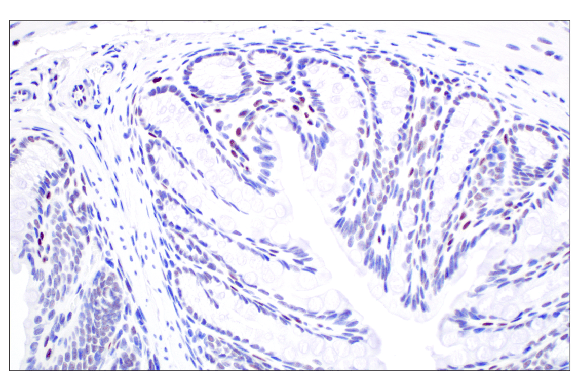
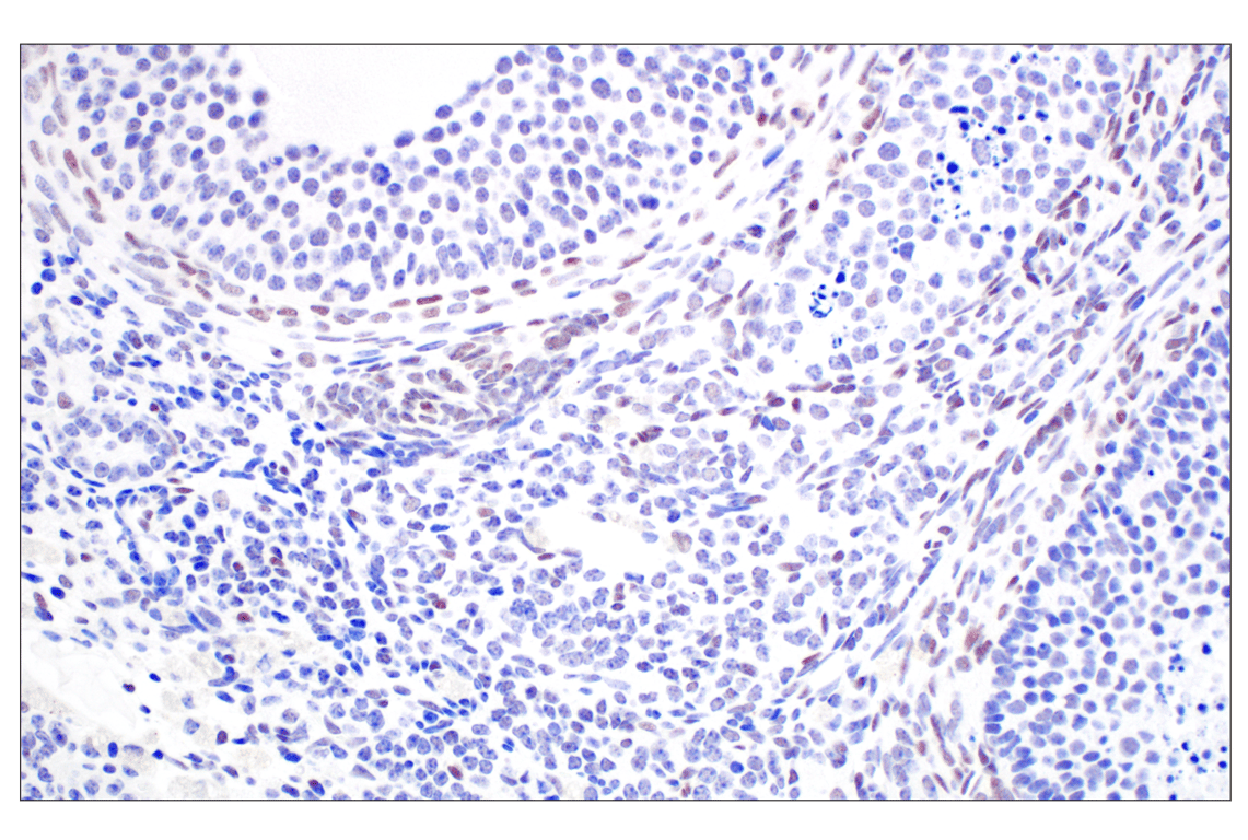
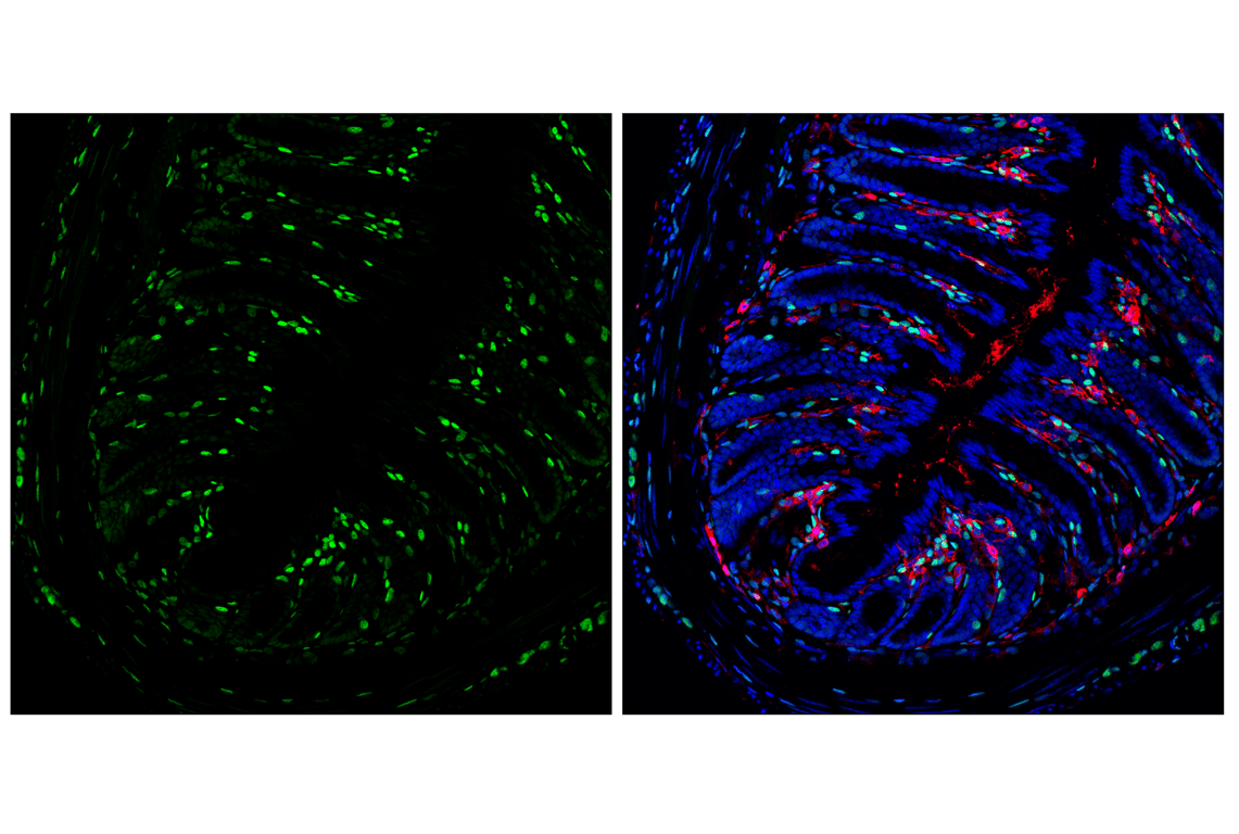
Immunohistochemical analysis of paraffin-embedded mouse colon using TCF4/ITF2 (E3X2Y) Rabbit mAb.

Immunohistochemistry Image 4: TCF4/ITF2 (E3X2Y) Rabbit mAb
Immunohistochemical analysis of paraffin-embedded mouse hippocampus using TCF4/ITF2 (E3X2Y) Rabbit mAb.
Immunohistochemistry Image 5: TCF4/ITF2 (E3X2Y) Rabbit mAb
Orders: 877-616-CELL (2355) [email protected] Support: 877-678-TECH (8324) [email protected] Web: cellsignal.com
For Research Use Only. Not for Use in Diagnostic Procedures.

Revision 3

CST Logo
Immunohistochemical analysis of paraffin-embedded mouse olfactory bulb using TCF4/ITF2 (E3X2Y) Rabbit mAb.
Immunohistochemistry Image 6: TCF4/ITF2 (E3X2Y) Rabbit mAb
Immunohistochemical analysis of paraffin-embedded mouse ovary using TCF4/ITF2 (E3X2Y) Rabbit mAb.
Immunohistochemistry Image 7: TCF4/ITF2 (E3X2Y) Rabbit mAb
Immunohistochemical analysis of paraffin-embedded mouse spleen using TCF4/ITF2 (E3X2Y) Rabbit mAb (left) or a TCF4/ITF2 Rabbit mAb (right). These two antibodies detect unique, non-overlapping epitopes on mouse TCF4/ITF2. The similar staining patterns obtained with both antibodies help to confirm the specificity of the staining.
Immunohistochemistry Image 8: TCF4/ITF2 (E3X2Y) Rabbit mAb
Orders: 877-616-CELL (2355) [email protected] Support: 877-678-TECH (8324) [email protected] Web: cellsignal.com
For Research Use Only. Not for Use in Diagnostic Procedures.

Revision 3

CST Logo
Immunohistochemical analysis of paraffin-embedded mouse spleen using TCF4/ITF2 (E3X2Y) Rabbit mAb (left) compared to concentration-matched Rabbit (DA1E) mAb IgG XP® Isotype Control #3900 (right).
Immunohistochemistry Image 9: TCF4/ITF2 (E3X2Y) Rabbit mAb
Immunohistochemical analysis of paraffin-embedded A20 cell pellet (left, positive) or EL4 cell pellet (right, negative) using TCF4/ITF2 (E3X2Y) Rabbit mAb.
Immunohistochemistry Image 10: TCF4/ITF2 (E3X2Y) Rabbit mAb
Confocal immunofluorescent analysis of fixed frozen mouse colon labeled with TCF4/ITF2 (E3X2Y) Rabbit mAb (green), CD45 (30-F11) Rat mAb #55307 (red), and ProLong Gold Antifade Reagent with DAPI #8961 (blue).
Immunofluorescence Image 1: TCF4/ITF2 (E3X2Y) Rabbit mAb
Orders: 877-616-CELL (2355) [email protected] Support: 877-678-TECH (8324) [email protected] Web: cellsignal.com
For Research Use Only. Not for Use in Diagnostic Procedures.

Revision 3

CST Logo
Confocal immunofluorescent analysis of fixed frozen mouse hippocampus labeled with TCF4/ITF2 (E3X2Y) Rabbit mAb (green) and ProLong Gold Antifade Reagent with DAPI #8961 (blue).
Immunofluorescence Image 2: TCF4/ITF2 (E3X2Y) Rabbit mAb
Confocal immunofluorescent analysis of fixed frozen mouse spleen labeled with TCF4/ITF2 (E3X2Y) Rabbit mAb (green), CD45 (30-F11) Rat mAb #55307 (red), and ProLong Gold Antifade Reagent with DAPI #8961 (blue).
Immunofluorescence Image 3: TCF4/ITF2 (E3X2Y) Rabbit mAb
Confocal immunofluorescent analysis of NCI-H520 cells (left, positive) and JAR cells (right, negative) using TCF4/ITF2 (E3X2Y) Rabbit mAb (green), DyLight 554 Phalloidin #13054 (red), and DAPI #4083 (blue).
Immunofluorescence Image 1: TCF4/ITF2 (E3X2Y) Rabbit mAb
Orders: 877-616-CELL (2355) [email protected] Support: 877-678-TECH (8324) [email protected] Web: cellsignal.com
For Research Use Only. Not for Use in Diagnostic Procedures.

Revision 3

CST Logo
Flow cytometric analysis of fixed/permeabilized Kasumi-1 cells (blue, negative) and KMS-11 cells (green, positive) using TCF4/ITF2 (E3X2Y) Rabbit mAb (solid lines) or concentration-matched Rabbit (DA1E) mAb IgG XP® Isotype Control #3900 (dashed lines). Anti-rabbit IgG F(ab')2 Fragment (Alexa Fluor® 488 Conjugate) #4412 was used as a secondary antibody.
Flow Cytometry Image 1: TCF4/ITF2 (E3X2Y) Rabbit mAb
Flow cytometric analysis of fixed/permeabilized EL4 cells (blue, low-expressing) and A20 cells (green, high-expressing) using TCF4/ITF2 (E3X2Y) Rabbit mAb (solid lines) or concentration-matched Rabbit (DA1E) mAb IgG XP® Isotype Control #3900 (dashed lines). Anti-rabbit IgG F(ab')2 Fragment (Alexa Fluor® 488 Conjugate) #4412 was used as a secondary antibody.
Flow Cytometry Image 2: TCF4/ITF2 (E3X2Y) Rabbit mAb
Orders: 877-616-CELL (2355) [email protected] Support: 877-678-TECH (8324) [email protected] Web: cellsignal.com
For Research Use Only. Not for Use in Diagnostic Procedures.