Revision 3

#36778

Store at -20C

CST Logo
Orders:

877-616-CELL (2355)

[email protected]

Support:

877-678-TECH (8324)

3 Trask Lane | Danvers | Massachusetts | 01923 | USA

For Research Use Only. Not for Use in Diagnostic Procedures.

Applications:
W, IP, IF-F, IF-IC, FC-FP, ChIP

Reactivity:
H M R

Sensitivity:
Endogenous

MW (kDa):
60-80

Source/Isotype:
Rabbit IgG

UniProt ID:
#Q96NM4, #O94900

Entrez-Gene Id:
84969, 9760

Product Usage Information

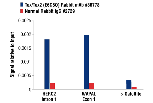
For optimal ChIP results, use 10 μl of antibody and 10 μg of chromatin (approximately 4 × 10⁶ cells) per IP. This antibody has been validated using SimpleChIP® Enzymatic Chromatin IP Kits.

Application Dilution
Western Blotting 1:1000
Immunoprecipitation 1:100
Immunofluorescence (Frozen) 1:100 - 1:400
Immunofluorescence (Immunocytochemistry) 1:100 - 1:400
Flow Cytometry (Fixed/Permeabilized) 1:200 - 1:800
Chromatin IP 1:50

Storage

Supplied in 10 mM sodium HEPES (pH 7.5), 150 mM NaCl, 100 µg/ml BSA, 50% glycerol and less than 0.02% sodium azide. Store at –20°C. Do not aliquot the antibody.

For a carrier free (BSA and azide free) version of this product see product #86380.

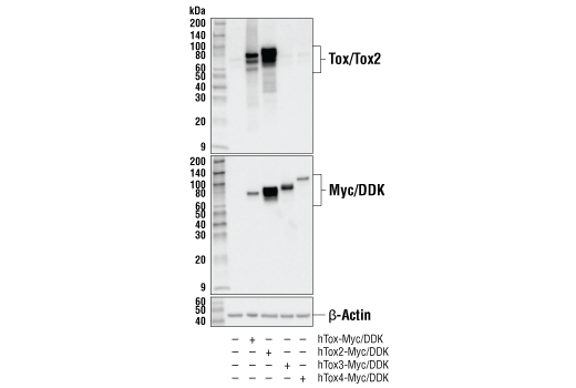
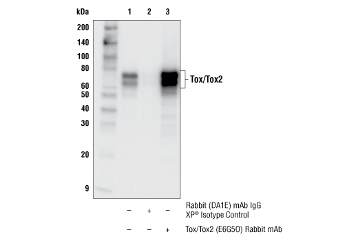
Specificity/Sensitivity

Tox/Tox2 (E6G5O) Rabbit mAb recognizes endogenous levels of total Tox and Tox2 proteins. This antibody does not cross-react with Tox3 or Tox4 proteins. This antibody cross-reacts with an unidentified protein of 50 kDa in some murine cell extracts.

Source / Purification

Monoclonal antibody is produced by immunizing animals with a synthetic peptide corresponding to residues surrounding Gln435 of human Tox protein.

Background

Thymocyte selection-associated high mobility group box protein (Tox) is a DNA-binding nuclear factor and member of the evolutionarily conserved high-motility group (HMG)-box superfamily. Tox also defines a small subfamily of proteins that include Tox2, Tox3, and Tox4, all of which are highly conserved in vertebrate species but have unique tissue expression patterns and functions (1,2).

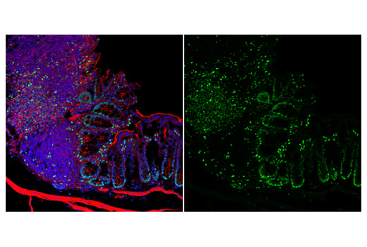
Tox plays a key role in T cell development in the thymus during positive selection (3-5). A study in Tox-deficient mice also revealed a requirement for Tox in CD4 T cell and NK cell lineage development, including NKT cells, FoxP3+ T regulatory T cells, and lymphoid tissue-inducer (LTi) cells (6-8). Although Tox expression is primarily restricted to developing immune cells in normal tissues, Tox is induced by high antigen stimulation during chronic viral infection or cancer, regulating T cell persistence and exhaustion (9-12). Tox has also been shown to be aberrantly expressed in cutaneous T cell lymphomas (13-14).

Species Reactivity

Species reactivity is determined by testing in at least one approved application (e.g., western blot).

Western Blot Buffer

IMPORTANT: For western blots, incubate membrane with diluted primary antibody in 5% w/v BSA, 1X TBS, 0.1% Tween® 20 at 4°C with gentle shaking, overnight.

Applications Key

W: Western Blotting IP: Immunoprecipitation IF-F: Immunofluorescence (Frozen) IF-IC: Immunofluorescence (Immunocytochemistry) FC-FP: Flow Cytometry (Fixed/Permeabilized) ChIP: Chromatin IP

Cross-Reactivity Key

H: Human M: Mouse R: Rat

Trademarks and Patents

Cell Signaling Technology is a trademark of Cell Signaling Technology, Inc.

Alexa Fluor is a registered trademark of Life Technologies Corporation.

KARPAS cell line source: Dr. Abraham Karpas at the University of Cambridge.

All other trademarks are the property of their respective owners. Visit cellsignal.com/trademarks for more information.

限制使用

除非 CST 的合法授书代表以书面形式书行明确同意,否书以下条款适用于 CST、其关书方或分书商提供的书品。 任何书充本条款或与本条款不同的客书条款和条件,除非书 CST 的合法授书代表以书面形式书独接受, 否书均被拒书,并且无效。

专品专有“专供研究使用”的专专或专似的专专声明, 且未专得美国食品和专品管理局或其他外国或国内专管机专专专任何用途的批准、准专或专可。客专不得将任何专品用于任何专断或治专目的, 或以任何不符合专专声明的方式使用专品。CST 专售或专可的专品提供专作专最专用专的客专,且专用于研专用途。将专品用于专断、专防或治专目的, 或专专售(专独或作专专成)或其他商专目的而专专专品,均需要 CST 的专独专可。客专:(a) 不得专独或与其他材料专合向任何第三方出售、专可、 出借、捐专或以其他方式专专或提供任何专品,或使用专品制造任何商专专品,(b) 不得复制、修改、逆向工程、反专专、 反专专专品或以其他方式专专专专专品的基专专专或技专,或使用专品开专任何与 CST 的专品或服专专争的专品或服专, (c) 不得更改或专除专品上的任何商专、商品名称、徽专、专利或版专声明或专专,(d) 只能根据 CST 的专品专售条款和任何适用文档使用专品 , (e) 专遵守客专与专品一起使用的任何第三方专品或服专的任何专可、服专条款或专似专专

Orders: 877-616-CELL (2355) [email protected] Support: 877-678-TECH (8324) [email protected] Web: cellsignal.com
For Research Use Only. Not for Use in Diagnostic Procedures.

Revision 3

Cell Signaling Technology Logo
Western blot analysis of extracts from various human cell lines using Tox/Tox2 (E6G5O) Rabbit mAb (upper) and β-Actin (D6A8) Rabbit mAb #8457 (lower).
Western Blotting Image 1: Tox/Tox2 (E6G5O) Rabbit mAb
Western blot analysis of extracts from human, mouse, and rat tissues and mouse cell lines using Tox/Tox2 (E6G5O) Rabbit mAb (upper) and β-Actin (D6A8) Rabbit mAb #8457 (lower).
Western Blotting Image 2: Tox/Tox2 (E6G5O) Rabbit mAb
Western blot analysis of extracts from 293T cells, mock transfected (-) or transfected (+) with constructs expressing Myc/DDK-tagged full-length human Tox protein (hTox-Myc/DDK), Myc/DDK-tagged full-length human Tox2 protein (hTox2-Myc/DDK), Myc/DDK-tagged full-length human Tox3 protein (hTox3-Myc/DDK), or Myc/DDK-tagged full-length human Tox4 protein (hTox4-Myc/DDK), using Tox/Tox2 (E6G5O) Rabbit mAb (upper), Myc-Tag (71D10) Rabbit mAb #2278 (middle), and β-Actin (D6A8) Rabbit mAb #8457 (lower).
Western Blotting Image 3: Tox/Tox2 (E6G5O) Rabbit mAb
Orders: 877-616-CELL (2355) [email protected] Support: 877-678-TECH (8324) [email protected] Web: cellsignal.com
For Research Use Only. Not for Use in Diagnostic Procedures.

Revision 3

Cell Signaling Technology Logo
Immunoprecipitation of Tox protein from SU-DHL-4 cell extracts. Lane 1 is 10% input, lane 2 is Rabbit (DA1E) mAb IgG XP® Isotype Control #3900, and lane 3 is Tox/Tox2 (E6G5O) Rabbit mAb. Western blot analysis was performed using Tox/Tox2 (E6G5O) Rabbit mAb.
Immunoprecipitation Image 1: Tox/Tox2 (E6G5O) Rabbit mAb
Confocal immunofluorescent analysis of mouse thymus (left) and pancreas (right) using Tox/Tox2 (E6G5O) Rabbit mAb (green). Actin filaments were labeled with DyLight™ 554 Phalloidin #13054 (red). Samples were mounted in ProLong® Gold Antifade Reagent with DAPI #8961 (blue).
Immunofluorescence Image 1: Tox/Tox2 (E6G5O) Rabbit mAb
Confocal immunofluorescent analysis of rat thymus (left) and pancreas (right) using Tox/Tox2 (E6G5O) Rabbit mAb (green). Actin filaments were labeled with DyLight™ 554 Phalloidin #13054 (red). Samples were mounted in ProLong® Gold Antifade Reagent with DAPI #8961 (blue).
Immunofluorescence Image 2: Tox/Tox2 (E6G5O) Rabbit mAb
Orders: 877-616-CELL (2355) [email protected] Support: 877-678-TECH (8324) [email protected] Web: cellsignal.com
For Research Use Only. Not for Use in Diagnostic Procedures.

Revision 3

Cell Signaling Technology Logo
Confocal immunofluorescent analysis of mouse colon using Tox/Tox2 (E6G5O) Rabbit mAb (green). Actin filaments were labeled with DyLight™ 554 Phalloidin #13054 (red). Samples were mounted in ProLong® Gold Antifade Reagent with DAPI #8961 (blue).
Immunofluorescence Image 3: Tox/Tox2 (E6G5O) Rabbit mAb
Confocal immunofluorescent analysis of SU-DHL-4 cells (left, positive) or A549 cells (right, negative) using Tox/Tox2 (E6G5O) Rabbit mAb (green). Actin filaments were labeled with DyLight™ 554 Phalloidin #13054 (red). Samples were mounted in ProLong® Gold Antifade Reagent with DAPI #8961 (blue).
Immunofluorescence Image 1: Tox/Tox2 (E6G5O) Rabbit mAb
Flow cytometric analysis of human peripheral blood mononuclear cells gated on lymphocytes co-stained with CD3 (UCHT1) Mouse mAb (APC Conjugate) #19881 using Tox/Tox2 (E6G5O) Rabbit mAb (right) compared to concentration matched

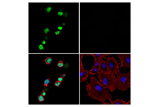
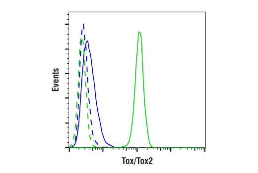
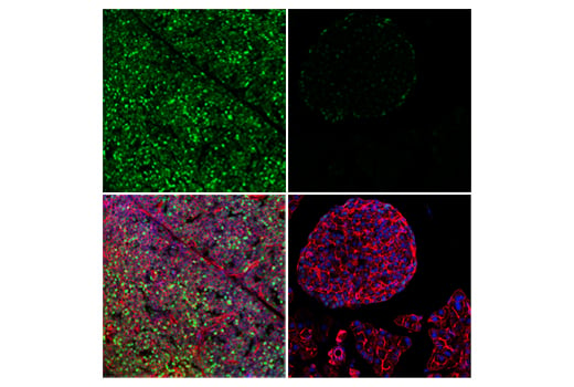
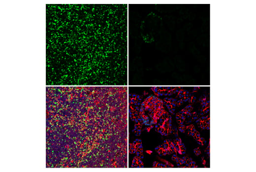
Rabbit (DA1E) mAb IgG XP® Isotype Control #3900 (left). Anti-rabbit IgG (H+L), F(ab')2 Fragment (Alexa Fluor® 488 Conjugate) #4412 was used as a secondary antibody.

Flow Cytometry Image 1: Tox/Tox2 (E6G5O) Rabbit mAb
Orders: 877-616-CELL (2355) [email protected] Support: 877-678-TECH (8324) [email protected] Web: cellsignal.com
For Research Use Only. Not for Use in Diagnostic Procedures.

Revision 3

Cell Signaling Technology Logo
Flow cytometric analysis of RPMI 8226 cells (blue, negative) and SU-DHL-4 cells (green, positive) using Tox/Tox2 (E6G5O) Rabbit mAb (solid lines) or a concentration-matched Rabbit (DA1E) mAb IgG XP® Isotype Control #3900 (dashed lines). Anti-rabbit IgG (H+L), F(ab')2 Fragment (Alexa Fluor® 488 Conjugate) #4412 was used as a secondary antibody.
Flow Cytometry Image 2: Tox/Tox2 (E6G5O) Rabbit mAb
Chromatin immunoprecipitations were performed with cross-linked chromatin from untreated SU-DHL-4 cells and either Tox/Tox2 (E6G5O) Rabbit mAb or Normal Rabbit IgG #2729 using SimpleChIP® Plus Enzymatic Chromatin IP Kit (Magnetic Beads) #9005. The enriched DNA was quantified by real-time PCR using human HERC2 intron 1 primers, human WAPAL exon 1 primers, and SimpleChIP® Human α Satellite Repeat Primers #4486. The amount of immunoprecipitated DNA in each sample is represented as signal relative to the total amount of input chromatin, which is equivalent to one.
Chromatin Immunoprecipitation Image 1: Tox/Tox2 (E6G5O) Rabbit mAb
Orders: 877-616-CELL (2355) [email protected] Support: 877-678-TECH (8324) [email protected] Web: cellsignal.com
For Research Use Only. Not for Use in Diagnostic Procedures.