Revision 1
#95052
Store at -20C
877-616-CELL (2355)
877-678-TECH (8324)
3 Trask Lane | Danvers | Massachusetts | 01923 | USA
For Research Use Only. Not for Use in Diagnostic Procedures.
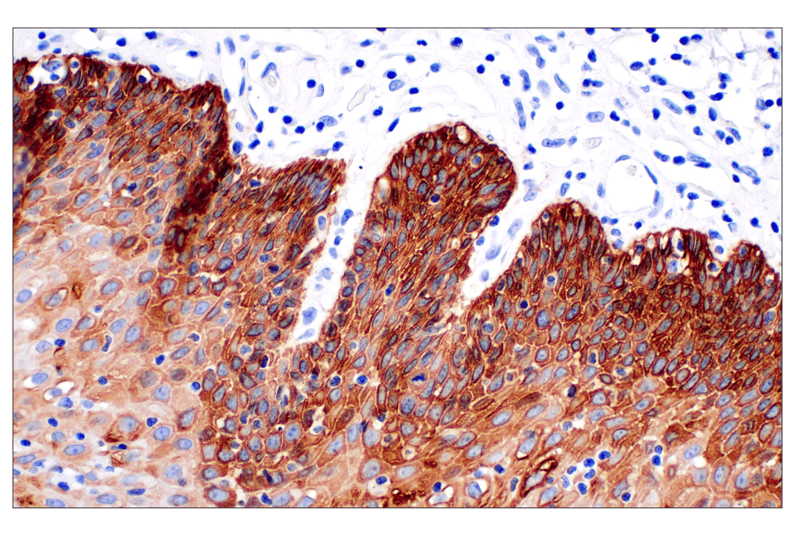
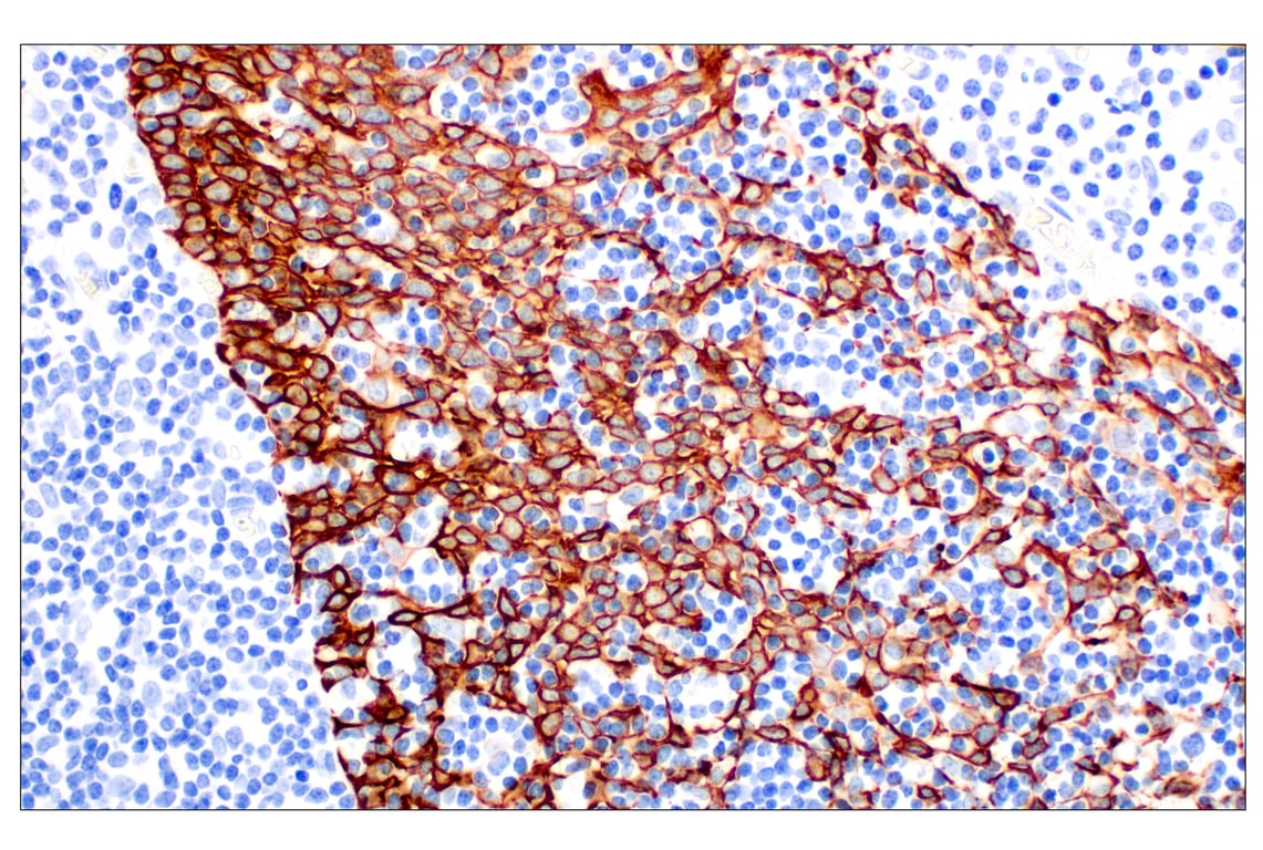
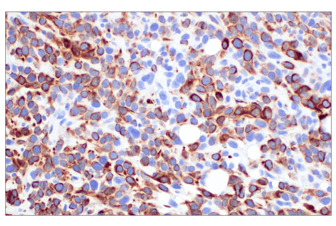
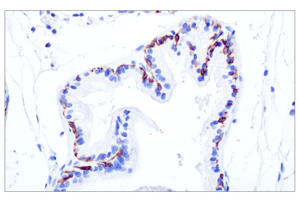
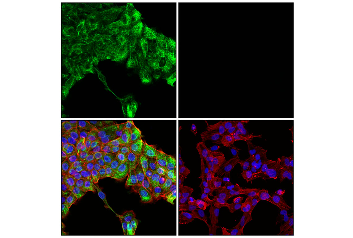
Applications:
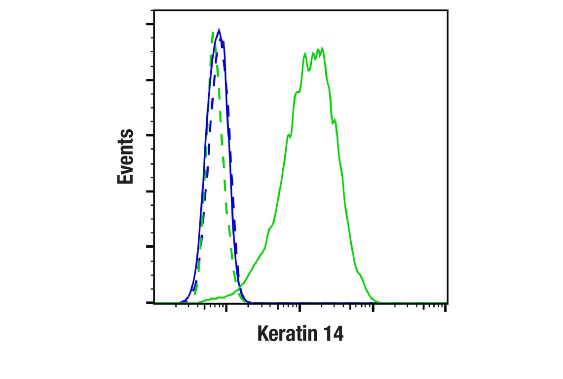
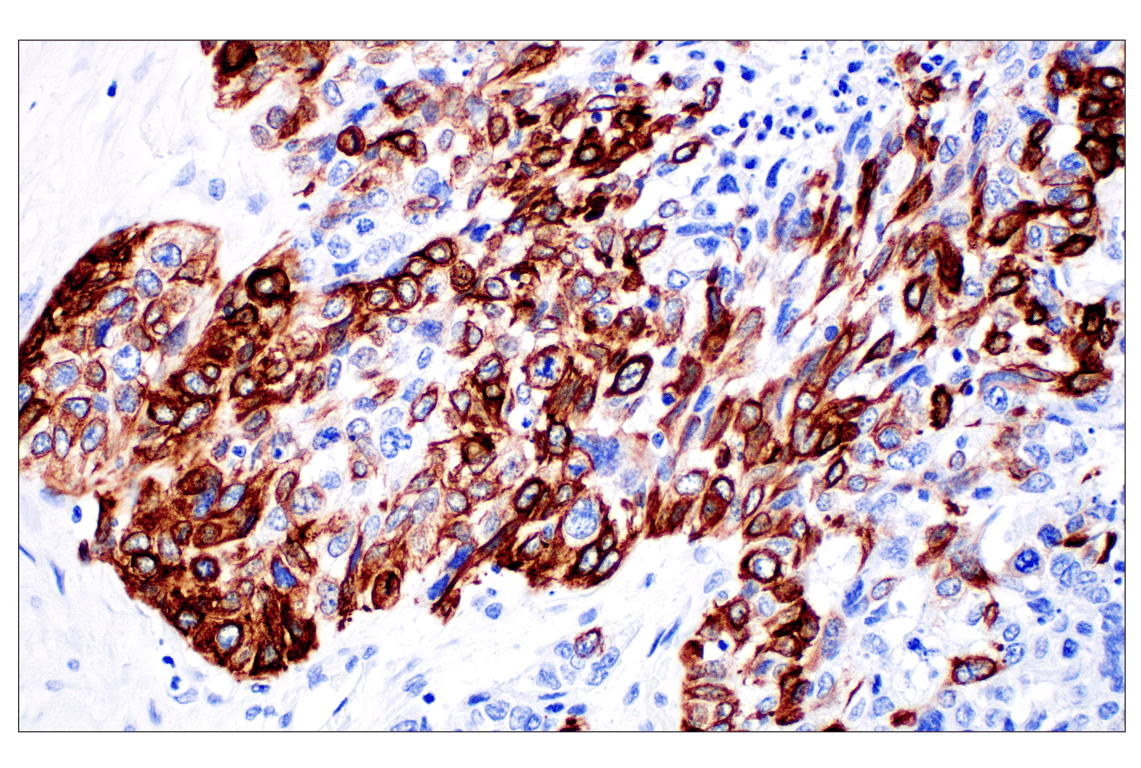
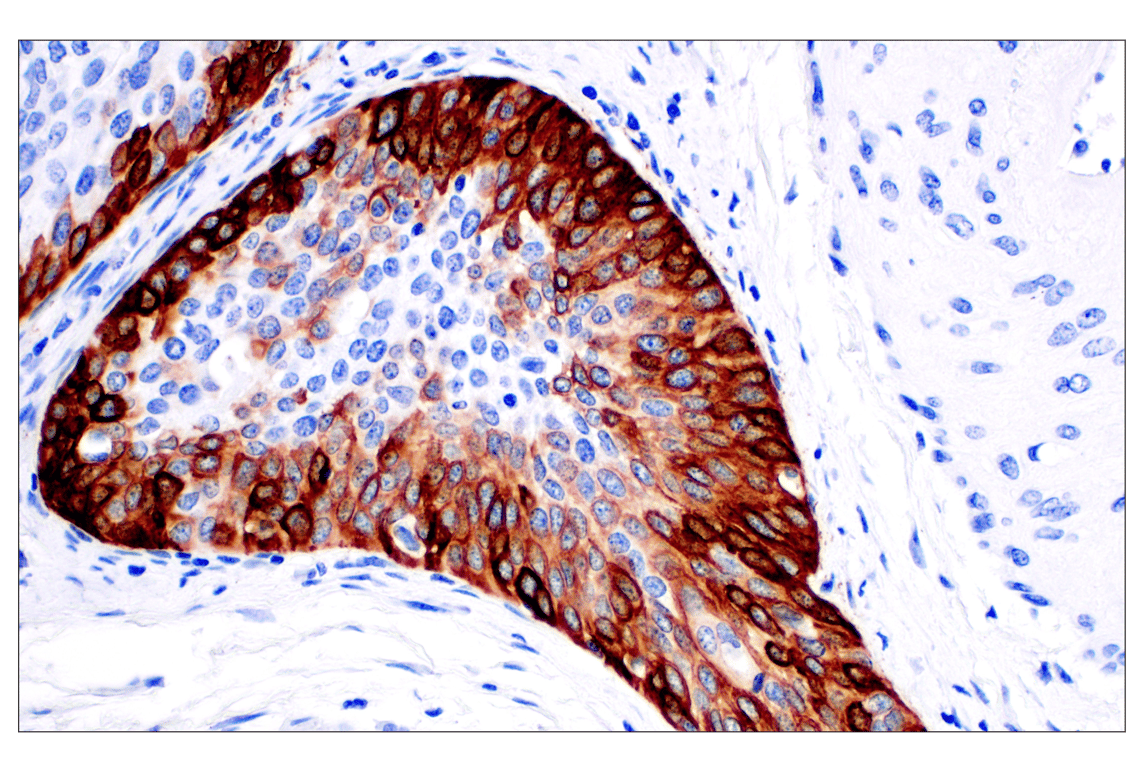
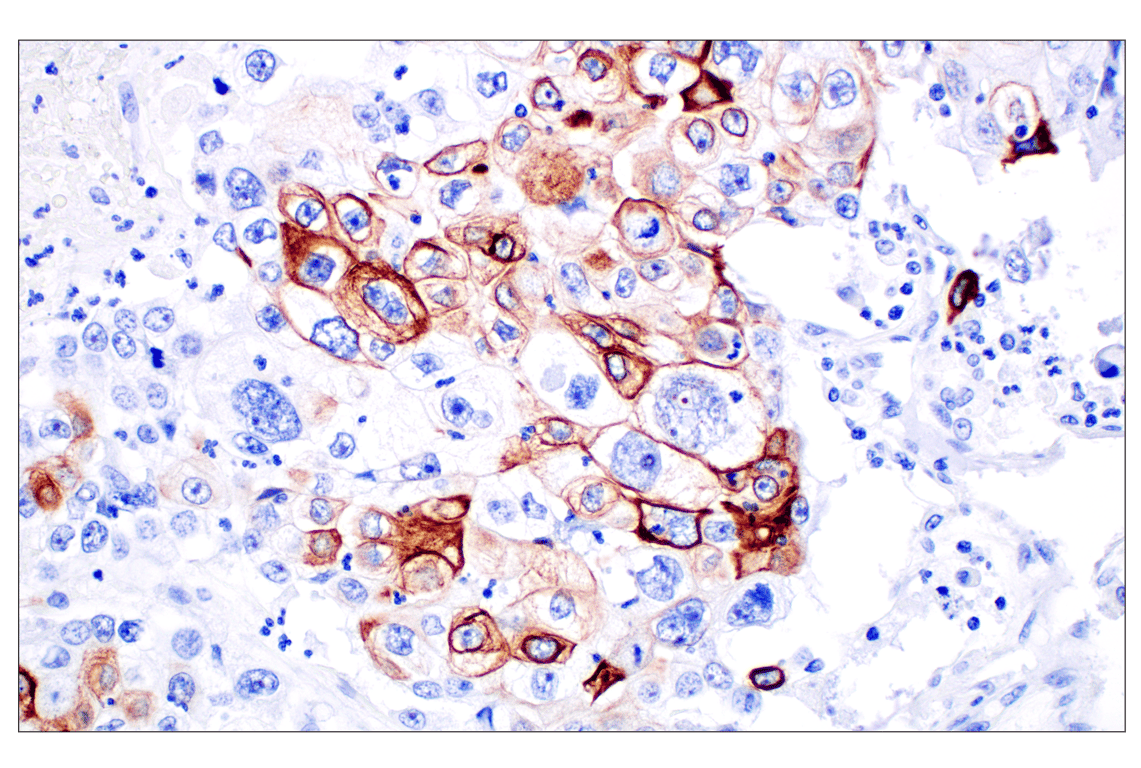
IHC-P, IF-F, IF-IC, FC-FP
Reactivity:
H M
Sensitivity:
Endogenous
Source/Isotype:
Rabbit IgG
UniProt ID:
#P02533
Entrez-Gene Id:
3861
Product Usage Information
This formulation is ideal for use with technologies requiring specialized or custom antibody labeling, including fluorophores, metals, lanthanides, and oligonucleotides. It is not recommended for ChIP, ChIP-seq, CUT&RUN or CUT&Tag assays. If you require a carrier free formulation for chromatin profiling, please contact us. Optimal dilutions/concentrations should be determined by the end user.
BSA and Azide Free antibodies are quality control tested by size exclusion chromatography (SEC) to determine antibody integrity.
Formulation
For standard formulation of this product see product #74956
Storage
Specificity/Sensitivity
Species predicted to react based on 100% sequence homology
Source / Purification
Background
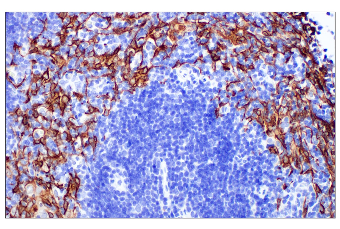
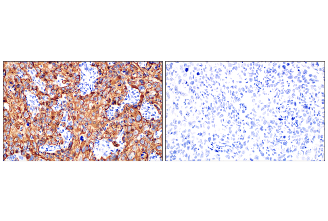
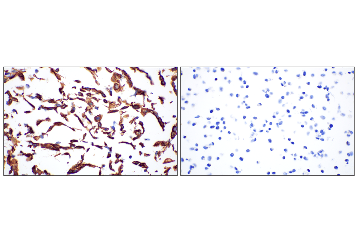
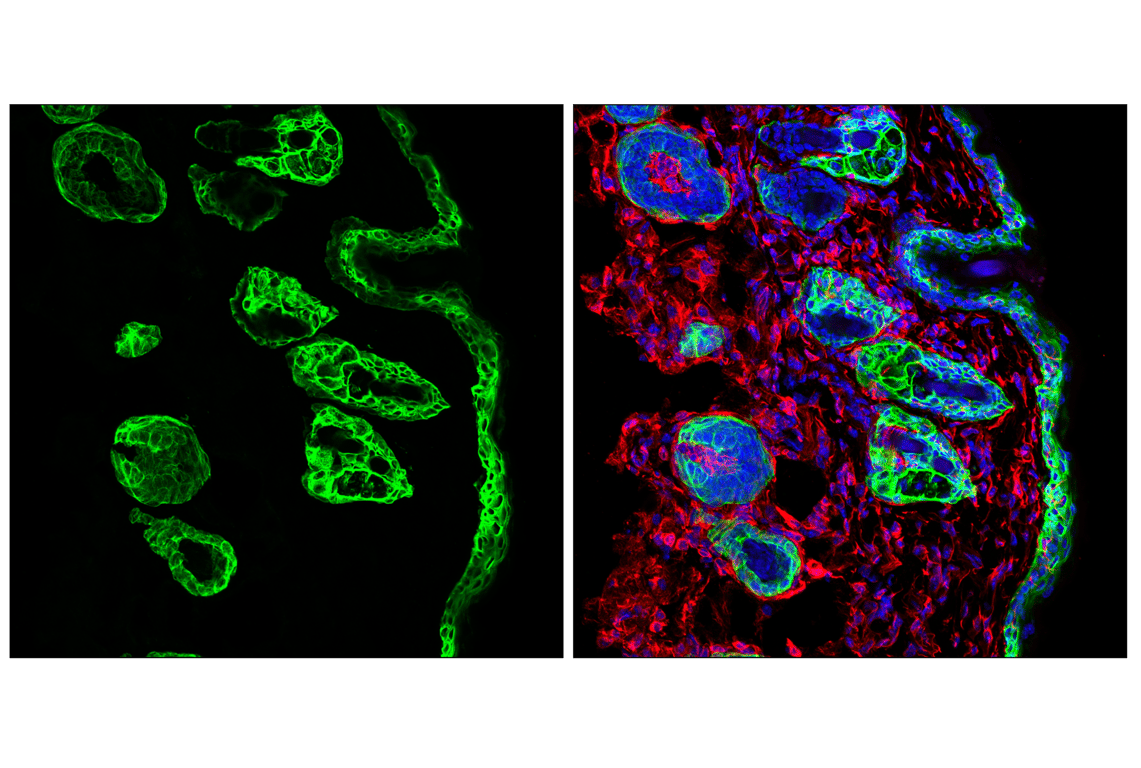
Dysregulation/mutations in keratin genes can lead to a variety of disorders affecting the skin, hair, nails, and other epithelial tissues (3). While expression of keratins can be variable, immunohistochemical staining of keratins is widely used to help in the identification and classification of epithelial tumors, and may also provide prognostic information.
Keratins 8 and 18 (K8/K18) are expressed in simple epithelia of normal tissue, as well as in adenocarcinomas of the breast, lung, ovary, and gastrointestinal tract. Keratin 17 is expressed in basal keratinocytes of stratified epithelia, hair follicles, and sebaceous glands. Onset of keratin 17 expression coincides with the definition of major epithelial lineages during skin development (4). Keratin 14 (K14) is expressed in basal cells of stratified epithelia, and in basal-like subtypes of breast cancer and squamous cell carcinomas. Keratin 19 (K19) is expressed in glandular epithelia, including the liver, gallbladder, and pancreas, as well as in adenocarcinomas of the breast, thyroid, and bile duct. Keratin 20 (K20) is expressed in gastrointestinal epithelium, urothelium, and Merkel cells in the skin, as well as in colorectal carcinomas and some urothelial carcinomas. Keratin 5/6 (K5/6) is expressed in basal cells of stratified epithelia, including the skin, prostate, and breast, as well as in basal-like breast cancers, squamous cell carcinomas, and some lung carcinomas. Keratin 7 (K7) is expressed in glandular epithelia, such as those in the lung, breast, and female reproductive tract, as well as in adenocarcinomas of the lung, breast, and ovary (5,6).
Keratins, particularly K8, K18, and K19, serve as biomarkers for identification of circulating tumor cells (CTCs) (5).
Post-translational modifications, including phosphorylation, acetylation, ubiquitylation, sumoylation, glycosylation, and transamidation, have been shown to affect the functions of keratins in normal and disease states (6). Understanding the molecular mechanisms underlying these PTMs may provide insights into cancer pathogenesis.
Background References
- Chang, L. and Goldman, R.D. (2004) Nat Rev Mol Cell Biol 5, 601-13.
- Schweizer, J. et al. (2006) J Cell Biol 174, 169-74.
- Sarma, A. (2022) Int J Biol Macromol 219, 395-413.
- McGowan, K.M. and Coulombe, P.A. (1998) J Cell Biol 143, 469-86.
- Werner, S. et al. (2020) Mol Aspects Med 72, 100817.
- Dmello, C. et al. (2019) J Biosci 44, 33.
Species Reactivity
Species reactivity is determined by testing in at least one approved application (e.g., western blot).
Applications Key
IHC-P: Immunohistochemistry (Paraffin) IF-F: Immunofluorescence (Frozen) IF-IC: Immunofluorescence (Immunocytochemistry) FC-FP: Flow Cytometry (Fixed/Permeabilized)
Cross-Reactivity Key
H: Human M: Mouse
Trademarks and Patents
Cell Signaling Technology is a trademark of Cell Signaling Technology, Inc.
All other trademarks are the property of their respective owners. Visit cellsignal.com/trademarks for more information.
限制使用
除非 CST 的合法授书代表以书面形式书行明确同意,否书以下条款适用于 CST、其关书方或分书商提供的书品。 任何书充本条款或与本条款不同的客书条款和条件,除非书 CST 的合法授书代表以书面形式书独接受, 否书均被拒书,并且无效。
专品专有“专供研究使用”的专专或专似的专专声明, 且未专得美国食品和专品管理局或其他外国或国内专管机专专专任何用途的批准、准专或专可。客专不得将任何专品用于任何专断或治专目的, 或以任何不符合专专声明的方式使用专品。CST 专售或专可的专品提供专作专最专用专的客专,且专用于研专用途。将专品用于专断、专防或治专目的, 或专专售(专独或作专专成)或其他商专目的而专专专品,均需要 CST 的专独专可。客专:(a) 不得专独或与其他材料专合向任何第三方出售、专可、 出借、捐专或以其他方式专专或提供任何专品,或使用专品制造任何商专专品,(b) 不得复制、修改、逆向工程、反专专、 反专专专品或以其他方式专专专专专品的基专专专或技专,或使用专品开专任何与 CST 的专品或服专专争的专品或服专, (c) 不得更改或专除专品上的任何商专、商品名称、徽专、专利或版专声明或专专,(d) 只能根据 CST 的专品专售条款和任何适用文档使用专品 , (e) 专遵守客专与专品一起使用的任何第三方专品或服专的任何专可、服专条款或专似专专
Revision 1
Revision 1
Revision 1
Revision 1
Revision 1