Revision 1
#83718
Store at -20C
Glutamine Metabolism Antibody Sampler Kit
1 Kit
(8 x 20 microliters)
877-616-CELL (2355)
877-678-TECH (8324)
3 Trask Lane | Danvers | Massachusetts | 01923 | USA
For Research Use Only. Not for Use in Diagnostic Procedures.
| Product Includes | Product # | Quantity | Mol. Wt | Isotype/Source |
|---|---|---|---|---|
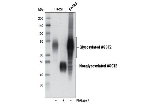
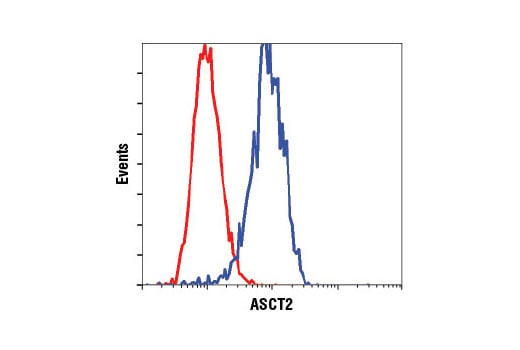
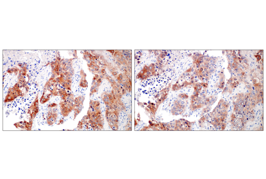
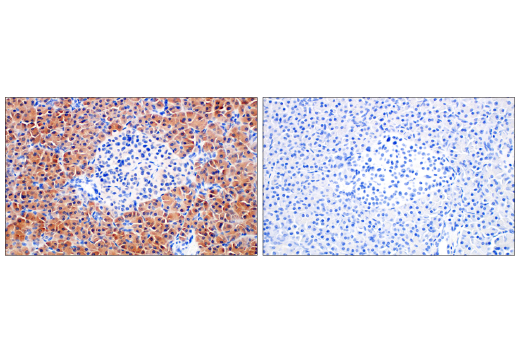
| ASCT2 (D7C12) Rabbit mAb | 8057 | 20 µl | 49, 75 kDa | Rabbit |
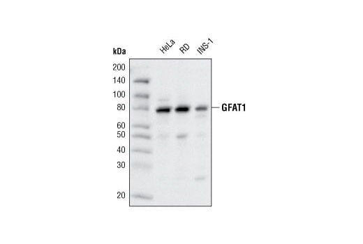
| GFAT1 (D12F4) Rabbit mAb | 5322 | 20 µl | 80 kDa | Rabbit IgG |
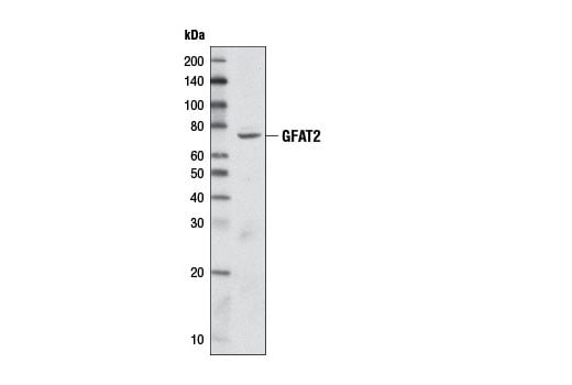
| GFAT2 (D40C7) Rabbit mAb | 6917 | 20 µl | 78 kDa | Rabbit IgG |
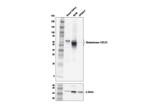
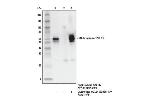
| Glutaminase-1/GLS1 (E9H6H) XP® Rabbit mAb | 56750 | 20 µl | 55-65 kDa | Rabbit IgG |
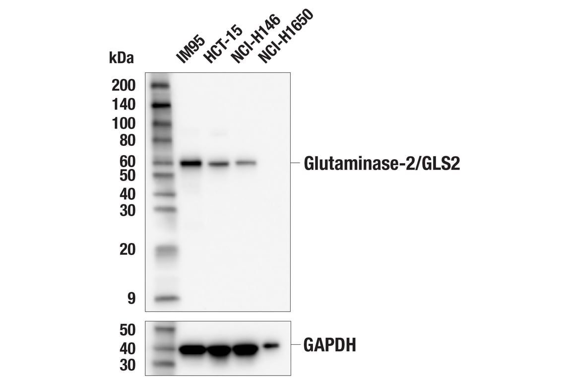
| Glutaminase-2/GLS2 (E9C7V) Rabbit mAb | 85934 | 20 µl | 60 kDa | Rabbit IgG |
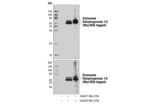
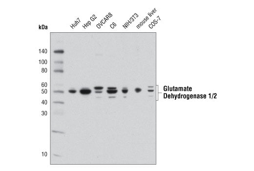
| Glutamate Dehydrogenase 1/2 (D9F7P) Rabbit mAb | 12793 | 20 µl | 52 kDa | Rabbit IgG |
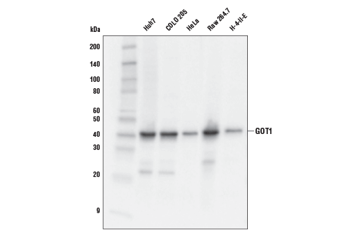
| GOT1 (E4A4O) Rabbit mAb | 34423 | 20 µl | 41 kDa | Rabbit IgG |
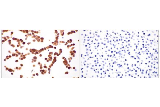
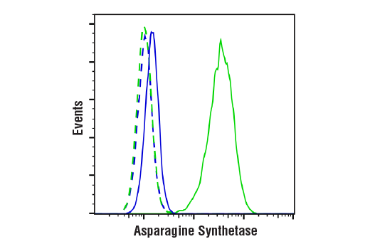
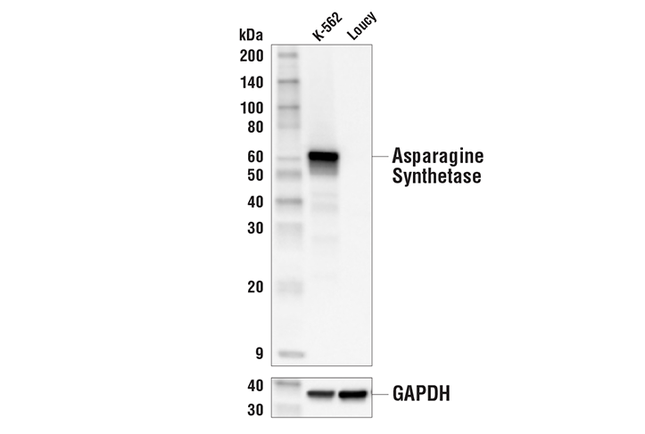
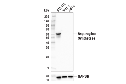
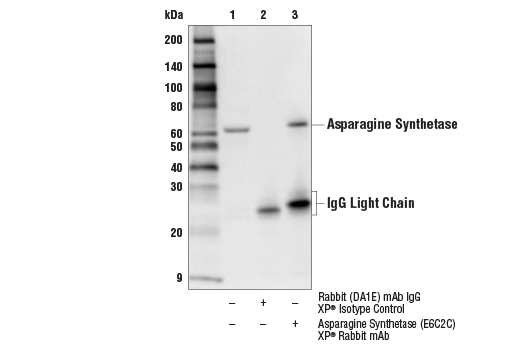
| Asparagine Synthetase (E6C2C) XP® Rabbit mAb | 92479 | 20 µl | 64 kDa | Rabbit IgG |
| Anti-rabbit IgG, HRP-linked Antibody | 7074 | 100 µl | Goat |
Please visit cellsignal.com for individual component applications, species cross-reactivity, dilutions, protocols, and additional product information.
Description
Storage
Background
GFAT1, glutamine:fructose-6-phosphate aminotransferase 1, is the rate-limiting enzyme of the hexosamine biosynthesis pathway (7). This enzyme catalyzes the conversion of fructose-6-phosphate and glutamine to glucosamine-6-phosphate and glutamate (8). The hexosamine biosynthesis pathway generates the building blocks for protein and lipid glycosylation (8). Furthermore, studies suggest that increased activity of this pathway is a contributing factor to hyperglycemia-induced insulin resistance (7,8). GFAT1 is more active in non-insulin-dependent diabetes mellitus (NIDDM) patients (9). Transgenic mice overexpressing this enzyme in skeletal muscle and adipose tissue show an insulin resistance phenotype (10,11). GFAT2, an isoenzyme of GFAT1, was later identified (12,13). Studies show that the regulation of GFAT2 is different from that of GFAT1, suggesting differential regulation of the hexosamine pathway in different tissues (13).
Glutaminase catalyzes the conversion of glutamine to glutamate, the first and rate-limiting step of glutaminolysis (14). Both kidney-type glutaminase (GLS1) and liver-type glutaminase (GLS2) are found in mammals (15). GLS1-mediated glutathione synthesis plays an essential role in redox homeostasis and contributes to increased survival of postimplantation bone cells preconditioned to the hypoxic and ischemic environment in the bone defect site (16). In addition, KEAP1–NRF2-mutant LUAD (KRAS-mutant lung adenocarcinoma) tumors are dependent on increased glutaminolysis (14). Furthermore, recent studies showed higher glutaminolysis and glucose production from glutamine in human primary hepatocytes with GLS2 gain-of-function missense mutations (17). These findings suggest GLS1 and GLS2 as potential targets in the therapy of bone regeneration and in the treatments of diseases such as cancer and hyperglycemia, respectively (14,16,17).
Glutamate dehydrogenase is a mitochondrial enzyme that catalyzes the oxidative deamination of glutamate to α-ketoglutarate through association with the cofactor nicotinamide adenine dinucleotide phosphate (18). Glutamate dehydrogenase is highly expressed in various tissues such as the liver, brain, kidney, heart, pancreas, ovaries, and testis. Two isoforms produced by two distinct genes are found in mammalian tissues. The GLUD1 gene is ubiquitously expressed (19), while the GLUD2 gene is specifically expressed in testicular tissues and astrocytes (20,21). Glutamate dehydrogenase links glutamate to the Krebs cycle, thereby playing a critical role in the regulation of energy homeostasis. Research studies have shown that changes in glutamate dehydrogenase activity in pancreatic β-cells can cause a hyperinsulinism syndrome (22).
Glutamate oxaloacetate transaminase 1 (GOT1) catalyzes the interconversion of aspartate and oxaloacetate (23).
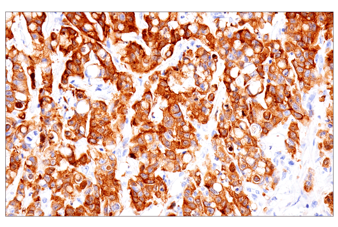
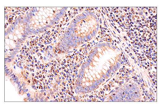
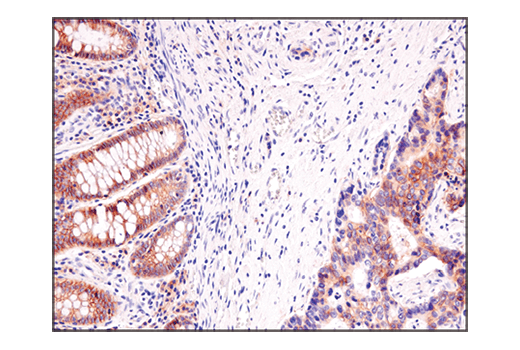
Asparagine synthetase (ASNS) catalyzes the synthesis of asparagine from aspartate and glutamine (24). In subsets of gastric and hepatic cancers, ASNS promoter hypermethylation correlates with low ASNS expression, sensitizing these cancers to the asparaginase treatment (25).
Background References
- Utsunomiya-Tate, N. et al. (1996) J Biol Chem 271, 14883-90.
- Bröer, S. (2008) Physiol Rev 88, 249-86.
- Bode, B.P. et al. (2002) Am J Physiol Gastrointest Liver Physiol 283, G1062-73.
- Fuchs, B.C. et al. (2007) Am J Physiol Cell Physiol 293, C55-63.
- Nicklin, P. et al. (2009) Cell 136, 521-34.
- Marin, M. et al. (2003) J Virol 77, 2936-45.
- Niimi, M. et al. (2001) J Hum Genet 46, 566-71.
- DeHaven, J.E. et al. (2001) Diabetes 50, 2419-24.
- Yki-Järvinen, H. et al. (1999) Life Sci 65, 215-23.
- Cooksey, R.C. et al. (1999) Endocrinology 140, 1151-7.
- Hebert, L.F. et al. (1996) J Clin Invest 98, 930-6.
- Oki, T. et al. (1999) Genomics 57, 227-34.
- Hu, Y. et al. (2004) J Biol Chem 279, 29988-93.
- Romero, R. et al. (2017) Nat Med 23, 1362-1368.
- Aledo, J.C. et al. (2000) Mamm Genome 11, 1107-10.
- Stegen, S. et al. (2016) Cell Metab 23, 265-79.
- Miller, R.A. et al. (2018) Nat Med 24, 518-524.
- Blumenthal, K.M. et al. (1975) J Biol Chem 250, 3644-54.
- Michaelidis, T.M. et al. (1993) Genomics 16, 150-60.
- Shashidharan, P. et al. (1997) J Neurochem 68, 1804-11.
- Zaganas, I. et al. (2012) Neurochem Int 61, 455-62.
- Karaca, M. et al. (2011) Neurochem Int 59, 510-7.
- Zhou, X. et al. (2018) BMC Cancer 18, 559.
- Zhang, J. et al. (2014) Mol Cell 56, 205-218.
- Li, H. et al. (2019) Nat Med 25, 850-860.
Trademarks and Patents
Cell Signaling Technology is a trademark of Cell Signaling Technology, Inc.
XP is a registered trademark of Cell Signaling Technology, Inc.
All other trademarks are the property of their respective owners. Visit cellsignal.com/trademarks for more information.
限制使用
除非 CST 的合法授书代表以书面形式书行明确同意,否书以下条款适用于 CST、其关书方或分书商提供的书品。 任何书充本条款或与本条款不同的客书条款和条件,除非书 CST 的合法授书代表以书面形式书独接受, 否书均被拒书,并且无效。
专品专有“专供研究使用”的专专或专似的专专声明, 且未专得美国食品和专品管理局或其他外国或国内专管机专专专任何用途的批准、准专或专可。客专不得将任何专品用于任何专断或治专目的, 或以任何不符合专专声明的方式使用专品。CST 专售或专可的专品提供专作专最专用专的客专,且专用于研专用途。将专品用于专断、专防或治专目的, 或专专售(专独或作专专成)或其他商专目的而专专专品,均需要 CST 的专独专可。客专:(a) 不得专独或与其他材料专合向任何第三方出售、专可、 出借、捐专或以其他方式专专或提供任何专品,或使用专品制造任何商专专品,(b) 不得复制、修改、逆向工程、反专专、 反专专专品或以其他方式专专专专专品的基专专专或技专,或使用专品开专任何与 CST 的专品或服专专争的专品或服专, (c) 不得更改或专除专品上的任何商专、商品名称、徽专、专利或版专声明或专专,(d) 只能根据 CST 的专品专售条款和任何适用文档使用专品 , (e) 专遵守客专与专品一起使用的任何第三方专品或服专的任何专可、服专条款或专似专专
Revision 1
Revision 1
Revision 1

Revision 1
Revision 1
Revision 1
Revision 1
Revision 1
Revision 1
Revision 1
Revision 1
Revision 1
Revision 1
Revision 1
Revision 1