Revision 4

#8873

Store at -20C

CST Logo
Orders:

877-616-CELL (2355)

[email protected]

Support:

877-678-TECH (8324)

3 Trask Lane | Danvers | Massachusetts | 01923 | USA

For Research Use Only. Not for Use in Diagnostic Procedures.

Applications:
W, W-S, IHC-P

Reactivity:
H M R

Sensitivity:
Endogenous

MW (kDa):
10, 57

Source/Isotype:
Rabbit IgG

UniProt ID:
#Q14790

Entrez-Gene Id:
841

Product Usage Information

Application Dilution
Western Blotting 1:1000
Simple Western™ 1:50 - 1:250
Immunohistochemistry (Paraffin) 1:50 - 1:200

Storage

Supplied in 10 mM sodium HEPES (pH 7.5), 150 mM NaCl, 100 µg/mL BSA, 50% glycerol, and less than 0.02% sodium azide. Store at –20°C. Do not aliquot the antibody.

For a carrier free (BSA and azide free) version of this product see product #46272.

Specificity/Sensitivity

Caspase-8 (F5K9P) Rabbit mAb recognizes endogenous levels of total caspase-8 protein, including the p10 subunit of the activated protein.

Source / Purification

Monoclonal antibody is produced by immunizing animals with a synthetic peptide corresponding to residues surrounding Pro394 of human caspase-8 protein.

Background

Apoptosis induced through the CD95 receptor (Fas/APO-1) and tumor necrosis factor receptor 1 (TNFR1) activates caspase-8 and leads to the release of the caspase-8 active fragments, p18 and p10 (1-3). Activated caspase-8 cleaves and activates downstream effector caspases such as caspase-1, -3, -6, and -7. Caspase-3 ultimately elicits the morphological hallmarks of apoptosis, including DNA fragmentation and cell shrinkage.

In addition to functioning as a key initiator caspase for extrinsic apoptosis, more recent studies have identified caspase-8 as a regulator of inflammatory necrotic cell death pathways such as necroptosis and pyroptosis (4,5). Activation of caspase-8 leads to cleavage of RIPK1 and RIPK3 to inhibit necroptosis (6,7). As a result, caspase-8 deficiency in mice, which is embryonic lethal, can be rescued by deletion of the necroptosis proteins RIPK3 or MLKL (8-11). Additionally, in some circumstances, caspase-8 is recruited to the inflammasome to trigger pyroptosis (12,13). Studies have also found that expression of an enzymatically inactive form of caspase-8 (C362S) causes embryonic lethality by inducing necroptosis and pyroptosis (14).

Species Reactivity

Species reactivity is determined by testing in at least one approved application (e.g., western blot).

Western Blot Buffer

IMPORTANT: For western blots, incubate membrane with diluted primary antibody in 5% w/v BSA, 1X TBS, 0.1% Tween® 20 at 4°C with gentle shaking, overnight.

Applications Key

W: Western Blotting W-S: Simple Western™ IHC-P: Immunohistochemistry (Paraffin)

Cross-Reactivity Key

H: Human M: Mouse R: Rat

Trademarks and Patents

Cell Signaling Technology is a trademark of Cell Signaling Technology, Inc.

All other trademarks are the property of their respective owners. Visit cellsignal.com/trademarks for more information.

限制使用

除非 CST 的合法授书代表以书面形式书行明确同意,否书以下条款适用于 CST、其关书方或分书商提供的书品。 任何书充本条款或与本条款不同的客书条款和条件,除非书 CST 的合法授书代表以书面形式书独接受, 否书均被拒书,并且无效。

专品专有“专供研究使用”的专专或专似的专专声明, 且未专得美国食品和专品管理局或其他外国或国内专管机专专专任何用途的批准、准专或专可。客专不得将任何专品用于任何专断或治专目的, 或以任何不符合专专声明的方式使用专品。CST 专售或专可的专品提供专作专最专用专的客专,且专用于研专用途。将专品用于专断、专防或治专目的, 或专专售(专独或作专专成)或其他商专目的而专专专品,均需要 CST 的专独专可。客专:(a) 不得专独或与其他材料专合向任何第三方出售、专可、 出借、捐专或以其他方式专专或提供任何专品,或使用专品制造任何商专专品,(b) 不得复制、修改、逆向工程、反专专、 反专专专品或以其他方式专专专专专品的基专专专或技专,或使用专品开专任何与 CST 的专品或服专专争的专品或服专, (c) 不得更改或专除专品上的任何商专、商品名称、徽专、专利或版专声明或专专,(d) 只能根据 CST 的专品专售条款和任何适用文档使用专品 , (e) 专遵守客专与专品一起使用的任何第三方专品或服专的任何专可、服专条款或专似专专

Orders: 877-616-CELL (2355) [email protected] Support: 877-678-TECH (8324) [email protected] Web: cellsignal.com
For Research Use Only. Not for Use in Diagnostic Procedures.

Revision 4

Cell Signaling Technology Logo
Western blot analysis of extracts from HCT 116 and CRISPR/Cas9 caspase-8 knockout (KO) HCT 116 cells, untreated (-) or treated with Staurosporine #9953 (1 μM, 4 hr; +), using Caspase-8 (F5K9P) Rabbit mAb (upper) or GAPDH (D16H11) XP® Rabbit mAb #5174 (lower).
Western Blotting Image 1: Caspase-8 (F5K9P) Rabbit mAb
Western blot analysis of extracts from THP-1 cells, untreated (-) or treated with Cycloheximide #2112 (CHX, 10 μg/mL, 18 hr) followed by Human Tumor Necrosis Factor-α (hTNF-α, 20 ng/mL, 4 hr; +), using Caspase-8 (F5K9P) Rabbit mAb (upper) or GAPDH (D16H11) XP® Rabbit mAb #5174 (lower).
Western Blotting Image 2: Caspase-8 (F5K9P) Rabbit mAb
Western blot analysis of extracts from WEHI-231 cells, untreated (-) or treated with Staurosporine #9953 (1 μM, 3 hr; +), using Caspase-8 (F5K9P) Rabbit mAb (upper) or GAPDH (D16H11) XP® Rabbit mAb #5174 (lower).
Western Blotting Image 3: Caspase-8 (F5K9P) Rabbit mAb
Orders: 877-616-CELL (2355) [email protected] Support: 877-678-TECH (8324) [email protected] Web: cellsignal.com
For Research Use Only. Not for Use in Diagnostic Procedures.

Revision 4

Cell Signaling Technology Logo
Western blot analysis of extracts from various cell lines using Caspase-8 (F5K9P) Rabbit mAb (upper) or GAPDH (D16H11) XP® Rabbit mAb #5174 (lower).
Western Blotting Image 4: Caspase-8 (F5K9P) Rabbit mAb
Western blot analysis of extracts from 293T cells, mock transfected (-) or transfected with a construct expressing Myc/DDK-tagged full-length human caspase-8 protein (hCasp8-Myc/DDK; +), using Caspase-8 (F5K9P) Rabbit mAb (upper), Myc-Tag (71D10) Rabbit mAb #2278 (middle), or GAPDH (D16H11) XP® Rabbit mAb #5174 (lower).
Western Blotting Image 5: Caspase-8 (F5K9P) Rabbit mAb
Simple Western™ analysis of lysates (1 mg/mL) from HCT 116 cells using Caspase-8 (F5K9P) Rabbit mAb #8873. The virtual lane view (left) shows the target band (as indicated) at 1:50 and 1:250 dilutions of primary antibody. The corresponding electropherogram view (right) plots chemiluminescence by molecular weight along the capillary at 1:50 (blue line) and 1:250 (green line) dilutions of primary antibody. This experiment was performed under reducing conditions on the Jess™ Simple Western instrument from ProteinSimple, a BioTechne brand, using the 12-230 kDa separation module.
Western Blotting Image 1: Caspase-8 (F5K9P) Rabbit mAb
Orders: 877-616-CELL (2355) [email protected] Support: 877-678-TECH (8324) [email protected] Web: cellsignal.com
For Research Use Only. Not for Use in Diagnostic Procedures.

Revision 4

Cell Signaling Technology Logo
Immunohistochemical analysis of paraffin-embedded human esophageal adenocarcinoma using Caspase-8 (F5K9P) Rabbit mAb.
Immunohistochemistry Image 1: Caspase-8 (F5K9P) Rabbit mAb

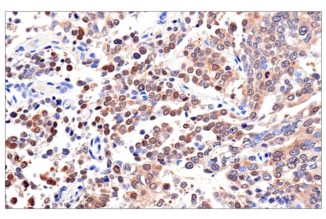
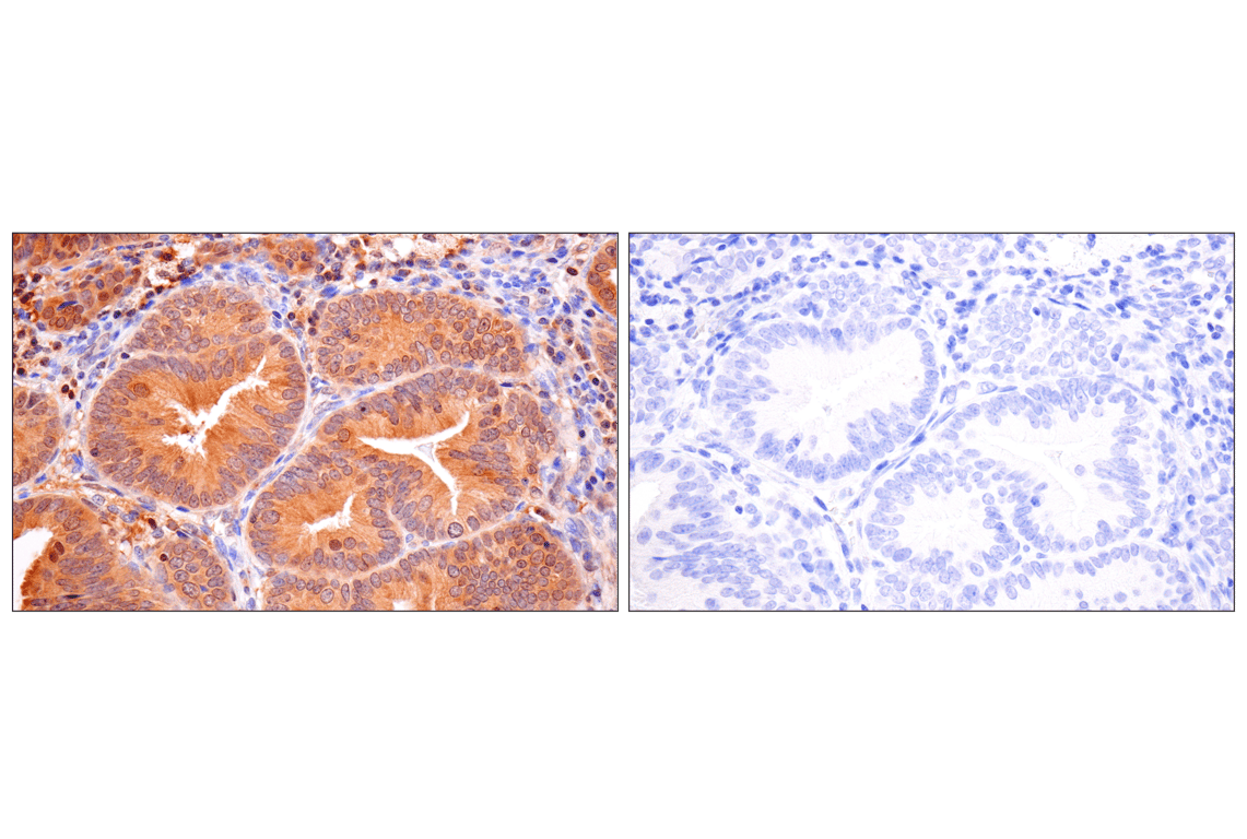
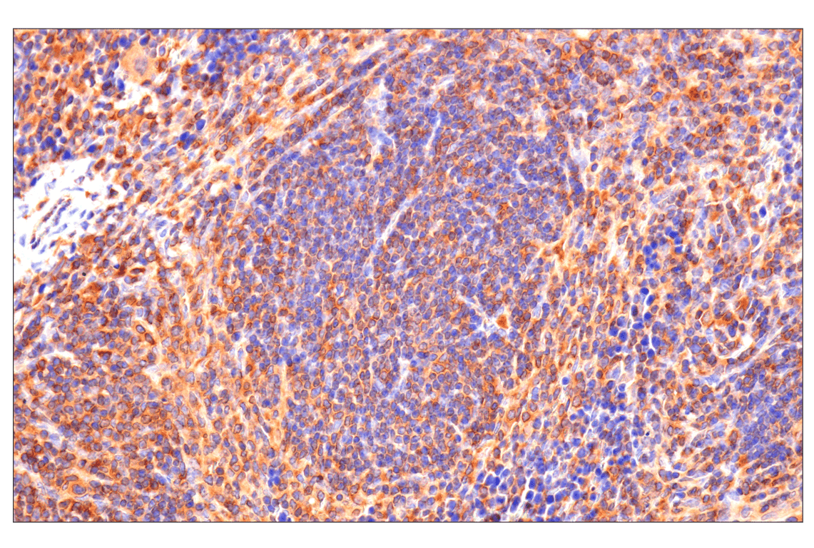
Immunohistochemical analysis of paraffin-embedded human granulosa cell tumor of the ovary using Caspase-8 (F5K9P) Rabbit mAb.

Immunohistochemistry Image 2: Caspase-8 (F5K9P) Rabbit mAb
Immunohistochemical analysis of paraffin-embedded human urothelial carcinoma (left), colon adenocarcinoma (middle), or normal kidney (right) using Caspase-8 (F5K9P) Rabbit mAb (top) or a Caspase-8 Rabbit mAb (bottom). These two antibodies detect unique, non-overlapping epitopes on human caspase-8. The similar staining patterns obtained with both antibodies help to confirm the specificity of the staining.
Immunohistochemistry Image 3: Caspase-8 (F5K9P) Rabbit mAb
Orders: 877-616-CELL (2355) [email protected] Support: 877-678-TECH (8324) [email protected] Web: cellsignal.com
For Research Use Only. Not for Use in Diagnostic Procedures.

Revision 4

Cell Signaling Technology Logo
Immunohistochemical analysis of paraffin-embedded 4T1 syngeneic tumor using Caspase-8 (F5K9P) Rabbit mAb.
Immunohistochemistry Image 4: Caspase-8 (F5K9P) Rabbit mAb

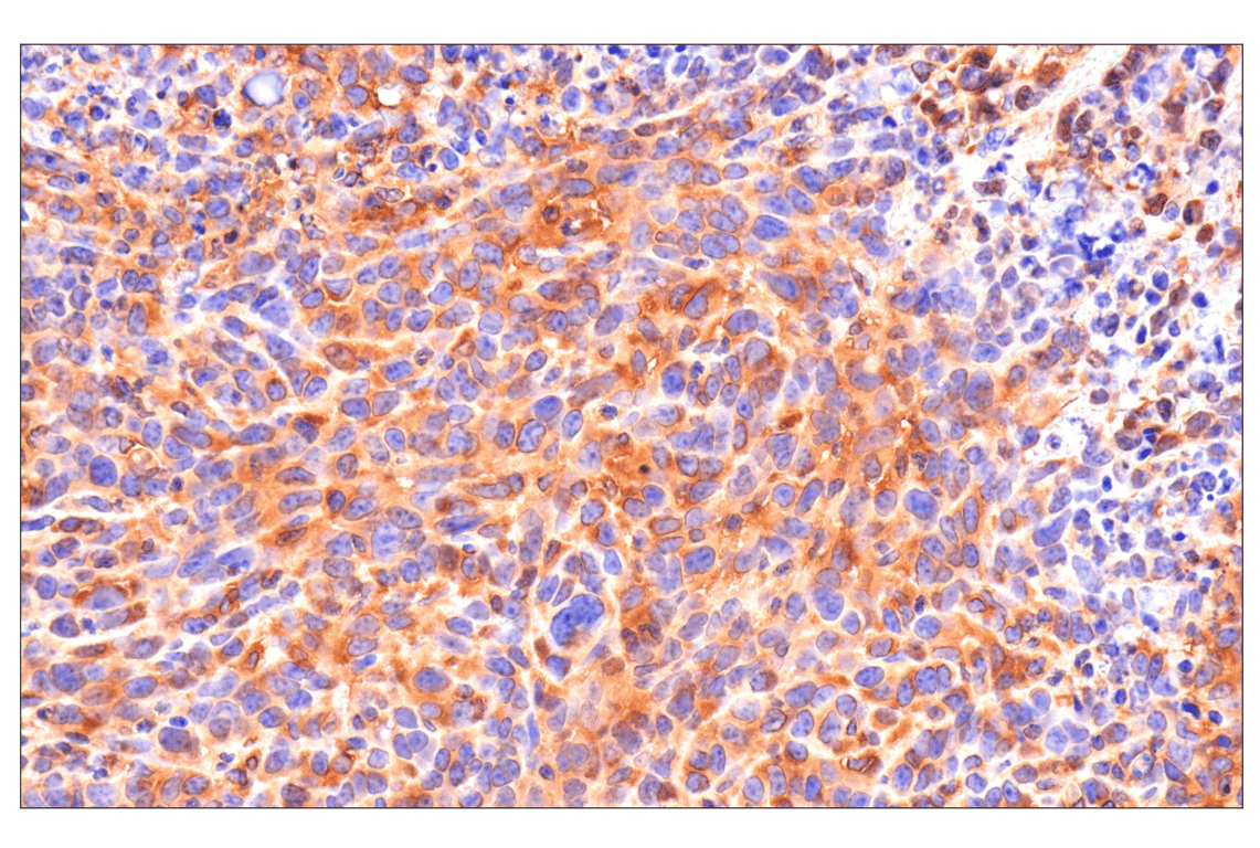
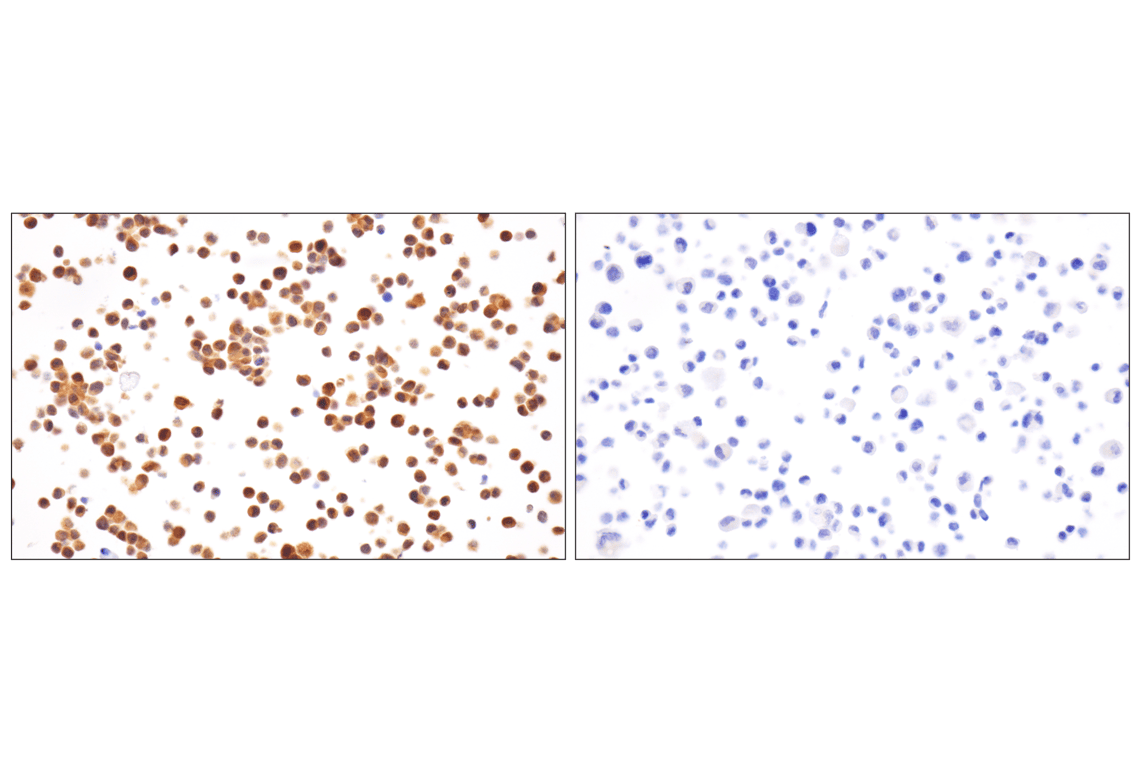
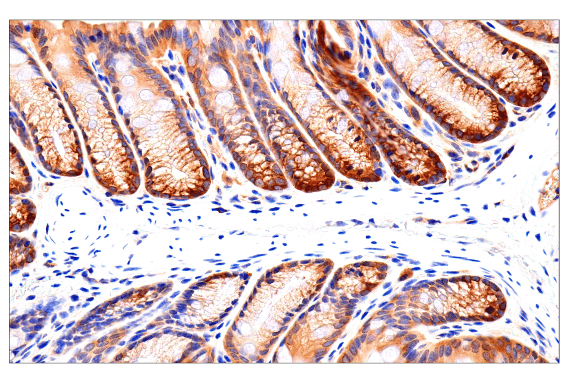
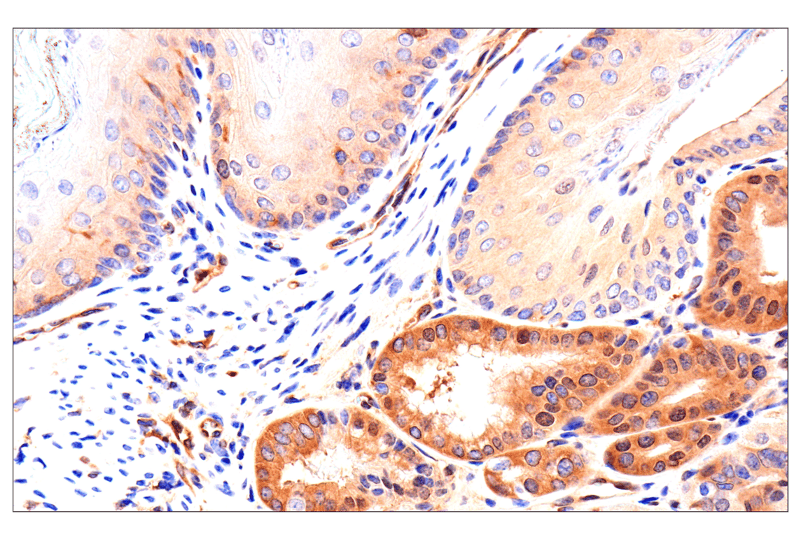
Immunohistochemical analysis of paraffin-embedded mouse colon using Caspase-8 (F5K9P) Rabbit mAb.

Immunohistochemistry Image 5: Caspase-8 (F5K9P) Rabbit mAb
Immunohistochemical analysis of paraffin-embedded mouse testis using Caspase-8 (F5K9P) Rabbit mAb.
Immunohistochemistry Image 6: Caspase-8 (F5K9P) Rabbit mAb
Orders: 877-616-CELL (2355) [email protected] Support: 877-678-TECH (8324) [email protected] Web: cellsignal.com
For Research Use Only. Not for Use in Diagnostic Procedures.

Revision 4

Cell Signaling Technology Logo
Immunohistochemical analysis of paraffin-embedded mouse spleen using Caspase-8 (F5K9P) Rabbit mAb.
Immunohistochemistry Image 7: Caspase-8 (F5K9P) Rabbit mAb
Immunohistochemical analysis of paraffin-embedded mouse thymus using Caspase-8 (F5K9P) Rabbit mAb.
Immunohistochemistry Image 8: Caspase-8 (F5K9P) Rabbit mAb
Immunohistochemical analysis of paraffin-embedded mouse forestomach using Caspase-8 (F5K9P) Rabbit mAb.
Immunohistochemistry Image 9: Caspase-8 (F5K9P) Rabbit mAb
Orders: 877-616-CELL (2355) [email protected] Support: 877-678-TECH (8324) [email protected] Web: cellsignal.com
For Research Use Only. Not for Use in Diagnostic Procedures.

Revision 4

Cell Signaling Technology Logo
Immunohistochemical analysis of paraffin-embedded human endometrioid adenocarcinoma using Caspase-8 (F5K9P) Rabbit mAb (left) compared to concentration-matched Rabbit (DA1E) mAb IgG XP® Isotype Control #3900 (right).
Immunohistochemistry Image 10: Caspase-8 (F5K9P) Rabbit mAb
Immunohistochemical analysis of paraffin-embedded HCT 116 cell pellets, control (left, positive) or CRISPR/Cas9 caspase-8 knockout (KO) (right, negative), using Caspase-8 (F5K9P) Rabbit mAb.
Immunohistochemistry Image 11: Caspase-8 (F5K9P) Rabbit mAb
Orders: 877-616-CELL (2355) [email protected] Support: 877-678-TECH (8324) [email protected] Web: cellsignal.com
For Research Use Only. Not for Use in Diagnostic Procedures.