Revision 1
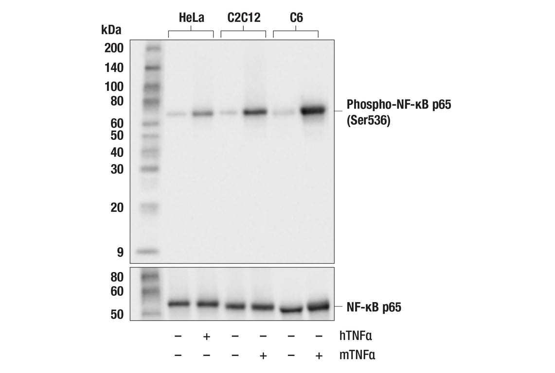
Western blot analysis of extracts from HeLa, C2C12, and C6 cells, untreated (-) or treated (+) as indicated with human TNF-α (hTNF⍺; 20ng/ml, 5min) or mouse TNF-α (mTNF⍺; 20ng/mL, 5min) using Phospho-NF-κB p65 (Ser536) (93H1) Rabbit mAb (upper) or NF-κB p65 (D14E12) XP® Rabbit mAb #8242 (lower). Phospho-NF-κB p65 is induced by human TNF-⍺ or mouse TNF-⍺ treatment as expected.
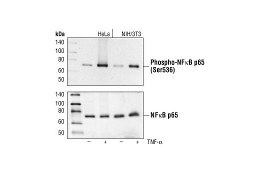
Western blot analysis of extracts from HeLa and NIH/3T3 cells, untreated or TNF-α treated (#2169, 20 ng/ml for 5 minutes), using Phospho-NF-κB p65 (Ser536) (93H1) Rabbit mAb (upper) or NF-κB p65 Antibody #3034 (lower).
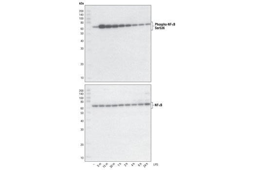
Western blot analysis of extracts from THP-1 cells, differentiated with TPA (#9905, 80 nM for 24h) and treated with 1 μg/ml LPS for the indicated times, using Phospho-NF-κB p65 (Ser536) (93H1) Rabbit mAb (upper) and NF-κB p65 (C22B4) Rabbit mAb #4764 (lower).
Orders: 877-616-CELL (2355) • [email protected] • Support: 877-678-TECH (8324) • [email protected] •
Web:
cellsignal.com For Research Use Only. Not for Use in Diagnostic Procedures.
Revision 1
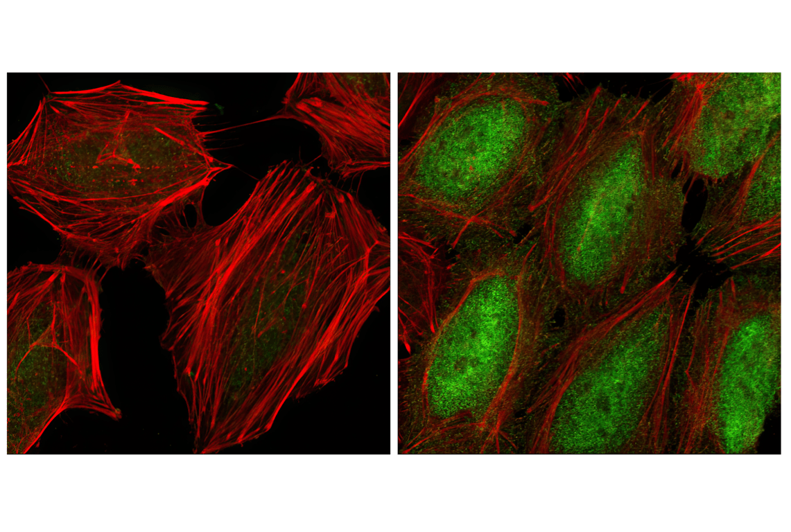
Confocal immunofluorescent analysis of HeLa cells, serum starved (left) or TNF-α treated (#8902 at 20 ng/ml for 20 min, right), using Phospho-NF-κB p65 (Ser536) (93H1) Rabbit mAb (green). Actin filaments have been labeled with Alexa Fluor® phalloidin 555 (red).
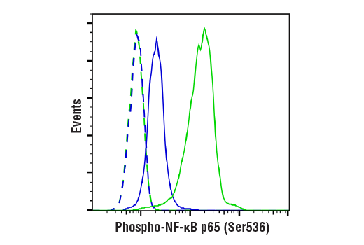
Flow cytometric analysis of HeLa cells, untreated (blue) or treated with Human Tumor Necrosis Factor-α (hTNF-α) #8902 and Calyculin A #9902 (20 ng/ml and 100 nM, 15 min; green), using Phospho-NF-κB p65 (Ser536) (93H1) Rabbit mAb (solid lines) or concentration-matched Rabbit (DA1E) mAb IgG XP® Isotype Control #3900 (dashed lines). Anti-rabbit IgG (H+L), F(ab')2 Fragment (Alexa Fluor® 488 Conjugate) #4412 was used as a secondary antibody.
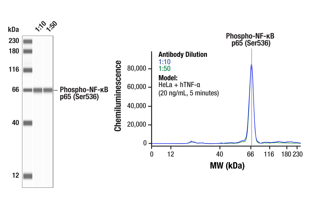
Simple Western™ analysis of lysates (1.0 mg/mL) from HeLa cells treated with hTNF-α (20 ng/mL, 5 minutes) using Phospho-NF-κB p65 (Ser536) (93H1) Rabbit mAb #3033. The virtual lane view (left) shows a single target band (as indicated) at 1:10 and 1:50 dilutions of primary antibody. The corresponding electropherogram view (right) plots chemiluminescence by molecular weight along the capillary at 1:10 (blue line) and 1:50 (green line) dilutions of primary antibody. This experiment was performed under reducing conditions on the Jess™ Simple Western instrument from ProteinSimple, a BioTechne brand, using the 12-230 kDa separation module.
Orders: 877-616-CELL (2355) • [email protected] • Support: 877-678-TECH (8324) • [email protected] •
Web:
cellsignal.com For Research Use Only. Not for Use in Diagnostic Procedures.
Revision 1
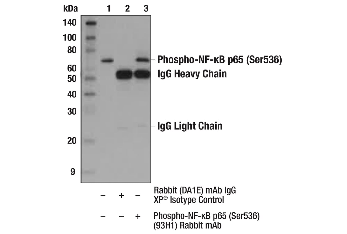
Immunoprecipitation of Phospho-NF-κB p65 (Ser536) from HeLa extracts treated with hTNF-α #8902 (20 ng/ml, 5 min). Lane 1 is 10% input, lane 2 is Rabbit (DA1E) mAb IgG XP® Isotype Control #3900, and lane 3 is Phospho-NF-κB p65 (Ser536) (93H1) Rabbit mAb. Western blot analysis was performed using Phospho-NF-κB p65 (Ser536) (93H1) Rabbit mAb. Anti-rabbit IgG, HRP-linked Antibody #7074 was used as a secondary antibody.
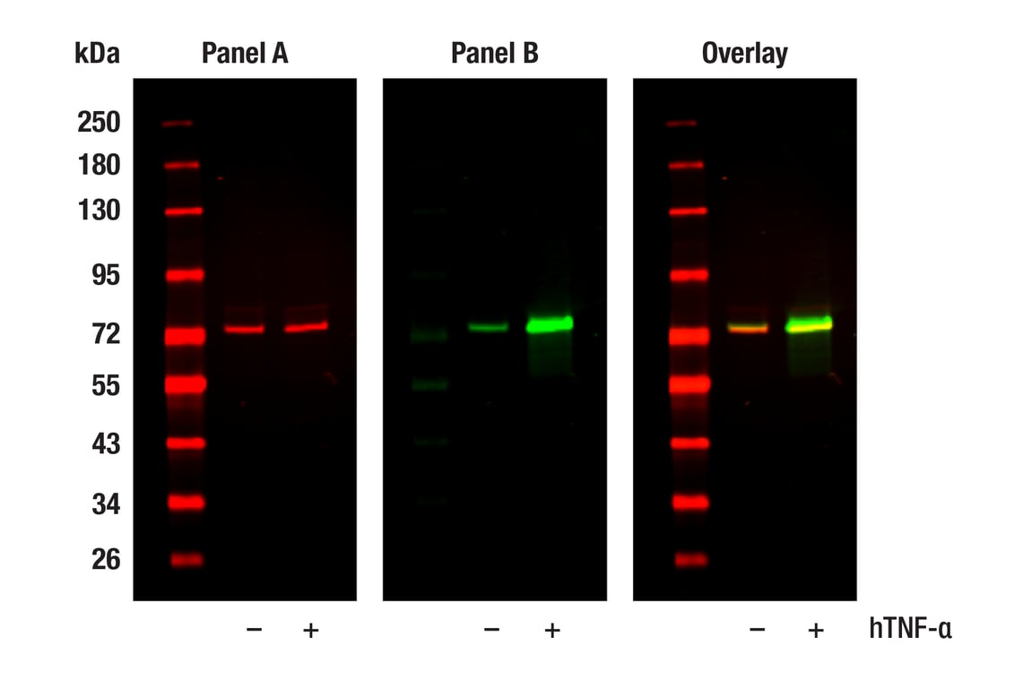
Western blot analysis of extracts from HeLa cells, untreated (-) or treated with hTNF-⍺ (20 ng/ml, 5 min; +), using NF-kB p65 (L8F6) Mouse mAb #6956 (Panel A) and Phospho-NF-kB p65 (Ser536) (93H1) Rabbit mAb #3033 (Panel B). Anti-mouse IgG (H+L) (DyLight 680 Conjugate) #5470 (red) and Anti-rabbit IgG (H+L) (DyLight 800 4X PEG Conjugate) #5151 (green) were used as secondary antibodies.
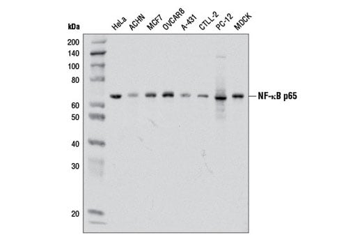
Western blot analysis of extracts from various cell lines using NF-κB p65 (D14E12) XP® Rabbit mAb.
Orders: 877-616-CELL (2355) • [email protected] • Support: 877-678-TECH (8324) • [email protected] •
Web:
cellsignal.com For Research Use Only. Not for Use in Diagnostic Procedures.
Revision 1
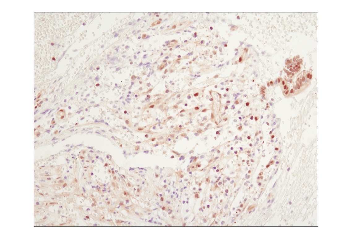
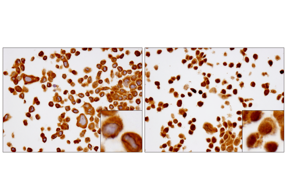
Immunohistochemical analysis of paraffin-embedded human chronic cholecystitis using NF-κB p65 (D14E12) XP® Rabbit mAb.
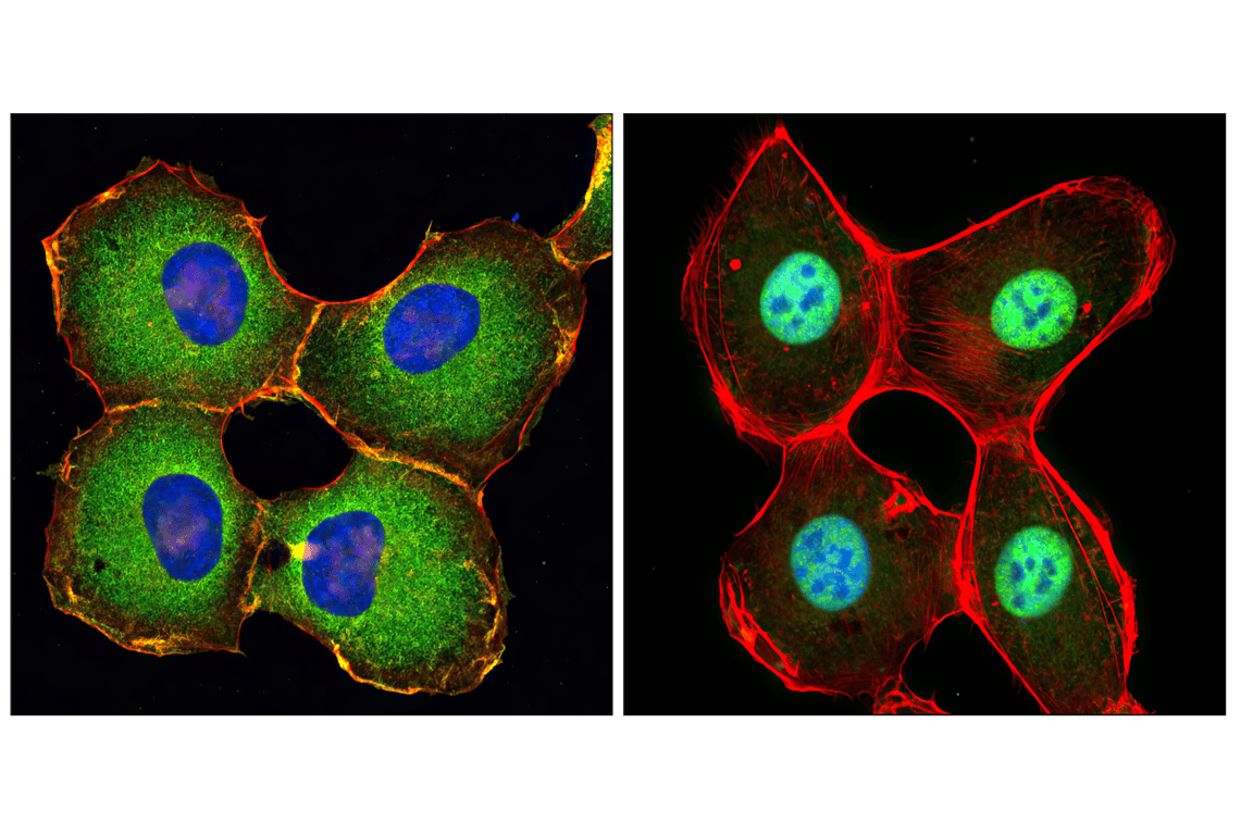
Confocal immunofluorescent analysis of HT-1080 cells, untreated (left) or treated with hTNF-α #8902 (20 ng/ml, 20 min) (right), using NF-κB p65 (D14E12) XP® Rabbit mAb (green). Actin filaments were labeled with DY-554 phalloidin (red). Blue pseudocolor = DRAQ5® #4084 (fluorescent DNA dye).
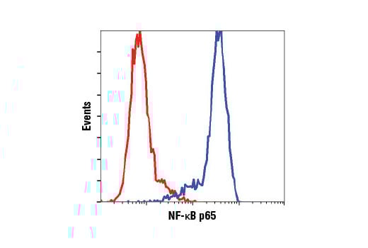
Flow cytometric analysis of HeLa cells using NF-κB p65 (D14E12) XP® Rabbit mAb (blue) compared to concentration matched Rabbit (DA1E) mAb IgG XP® Isotype Control #3900 (red).
Orders: 877-616-CELL (2355) • [email protected] • Support: 877-678-TECH (8324) • [email protected] •
Web:
cellsignal.com For Research Use Only. Not for Use in Diagnostic Procedures.
Revision 1
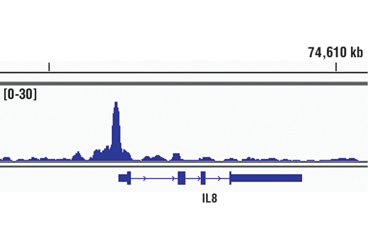
Chromatin immunoprecipitations were performed with cross-linked chromatin from HeLa cells treated with hTNF-α #8902 (30 ng/ml, 1 hr) and NF-κB p65 (D14E12) XP® Rabbit mAb, using SimpleChIP® Enzymatic Chromatin IP Kit (Magnetic Beads) #9005. DNA Libraries were prepared using DNA Library Prep Kit for Illumina® (ChIP-seq, CUT&RUN) #56795. The figure shows binding across IL-8, a known target gene of NFκB (see additional figure containing ChIP-qPCR data). For additional ChIP-seq tracks, please download the product datasheet.
Immunohistochemical analysis using NF-κB p65 (D14E12) XP® Rabbit mAb on SignalSlide® NF-κB p65 IHC Controls #12873 (paraffin-embedded HCT116 cells, untreated (left) or treated with hTNF-α #8902 (right)).
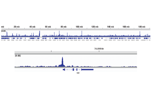
Chromatin immunoprecipitations were performed with cross-linked chromatin from HeLa cells treated with hTNF-α #8902 (30 ng/ml, 1 hr) and NF-κB p65 (D14E12) XP® Rabbit mAb, using SimpleChIP® Enzymatic Chromatin IP Kit (Magnetic Beads) #9005. DNA Libraries were prepared using DNA Library Prep Kit for Illumina® (ChIP-seq, CUT&RUN) #56795. The figure shows binding across chromosome 4 (upper), including IL-8 (lower), a known target gene of NFκB (see additional figure containing ChIP-qPCR data).
Orders: 877-616-CELL (2355) • [email protected] • Support: 877-678-TECH (8324) • [email protected] •
Web:
cellsignal.com For Research Use Only. Not for Use in Diagnostic Procedures.
Revision 1
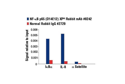
Chromatin immunoprecipitations were performed with cross-linked chromatin from HeLa cells treated with hTNF-α #8902 (30 ng/ml, 1 hr) and either NF-κB p65 (D14E12) XP® Rabbit mAb or Normal Rabbit IgG #2729 using SimpleChIP® Enzymatic Chromatin IP Kit (Magnetic Beads) #9003. The enriched DNA was quantified by Real-Time PCR using SimpleChIP® Human IκBα Promoter Primers #5552, human IL-8 promoter primers, and SimpleChIP® Human α Satellite Repeat Primers #4486. The amount of immunoprecipitated DNA in each sample is represented as signal relative to the total amount of input chromatin, which is equivalent to one.
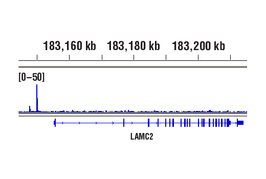
CUT&RUN was performed with HeLa cells treated with hTNF-α #8902 (30 ng/ml, 1 hr) and NF-κB p65 (D14E12) XP® Rabbit mAb, using CUT&RUN Assay Kit #86652. DNA Libraries were prepared using DNA Library Prep Kit for Illumina® (ChIP-seq, CUT&RUN) #56795. The figure shows binding across LAMC2, a known target gene of NF-κB p65 (see additional figure containing CUT&RUN-qPCR data).
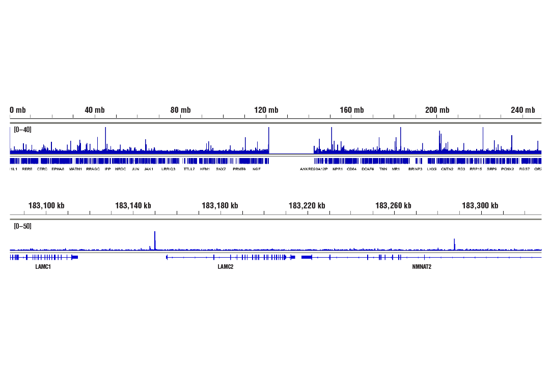
CUT&RUN was performed with HeLa cells treated with hTNF-α #8902 (30 ng/ml, 1 hr) and NF-κB p65 (D14E12) XP® Rabbit mAb, using CUT&RUN Assay Kit #86652. DNA Libraries were prepared using DNA Library Prep Kit for Illumina® (ChIP-seq, CUT&RUN) #56795. The figures show binding across chromosome 1 (upper), including LAMC2 (lower), a known target gene of NF-κB p65 (see additional figure containing CUT&RUN-qPCR data).
Orders: 877-616-CELL (2355) • [email protected] • Support: 877-678-TECH (8324) • [email protected] •
Web:
cellsignal.com For Research Use Only. Not for Use in Diagnostic Procedures.
Revision 1
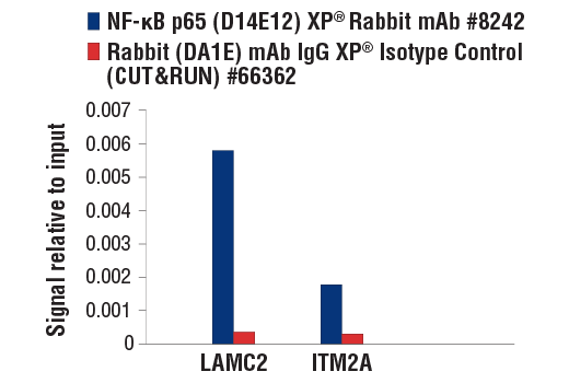
CUT&RUN was performed with HeLa cells treated with hTNF-α #8902 (30 ng/ml, 1 hr) and either NF-κB p65 (D14E12) XP® Rabbit mAb or Rabbit (DA1E) mAb IgG XP® Isotype Control (CUT&RUN) #66362, using CUT&RUN Assay Kit #86652. The enriched DNA was quantified by real-time PCR using human LAMC2 upstream primers, and human ITM2A upstream primers. The amount of immunoprecipitated DNA in each sample is represented as signal relative to the total amount of input chromatin, which is equivalent to one.
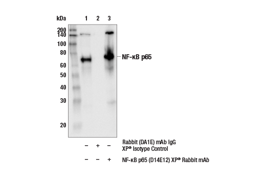
Immunoprecipitation of NF-kB p65 from CHO cell extracts. Lane 1 is 10% input, lane 2 is precipitated with Rabbit (DA1E) mAb IgG XP® Isotype Control #3900, and lane 3 is NF-κB p65 (D14E12) XP® Rabbit mAb, #8242. Western blot was performed using NF-κB p65 (L8F6) Mouse mAb, #6956.
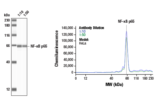
Simple Western™ analysis of lysates (1 mg/mL) from HeLa cells using NF-κB p65 (D14E12) XP® Rabbit mAb #8242. The virtual lane view (left) shows a single target band (as indicated) at 1:10 and 1:50 dilutions of primary antibody. The corresponding electropherogram view (right) plots chemiluminescence by molecular weight along the capillary at 1:10 (blue line) and 1:50 (green line) dilutions of primary antibody. This experiment was performed under reducing conditions on the Jess™ Simple Western instrument from ProteinSimple, a BioTechne brand, using the 12-230 kDa separation module.
Orders: 877-616-CELL (2355) • [email protected] • Support: 877-678-TECH (8324) • [email protected] •
Web:
cellsignal.com For Research Use Only. Not for Use in Diagnostic Procedures.