Revision 1
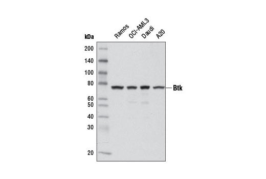
Western blot analysis of extracts from various cell lines using Btk (D3H5) Rabbit mAb.
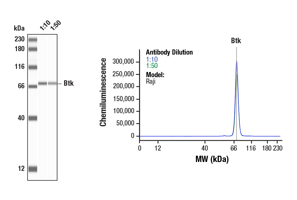
Simple Western™ analysis of lysates (0.1 mg/mL) from Raji cells using Btk (D3H5) Rabbit mAb #8547. The virtual lane view (left) shows a single target band (as indicated) at 1:10 and 1:50 dilutions of primary antibody. The corresponding electropherogram view (right) plots chemiluminescence by molecular weight along the capillary at 1:10 (blue line) and 1:50 (green line) dilutions of primary antibody. This experiment was performed under reducing conditions on the Jess™ Simple Western instrument from ProteinSimple, a BioTechne brand, using the 12-230 kDa separation module.
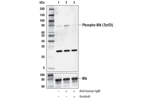
Western blot analysis of extracts from Daudi cells, serum-starved overnight, then vehicle-treated (lane 1), treated with anti-human IgM (12 μg/ml, 10 min; lane 2), or pre-treated with Ibrutinib #16483 (1 μM, 60 min) prior to anti-IgM treatment (lane 3), using Phospho-Btk (Tyr23) (D1D2Z) Rabbit mAb (upper) or Btk (D6H5) Rabbit mAb #8547 (lower).
Orders: 877-616-CELL (2355) • [email protected] • Support: 877-678-TECH (8324) • [email protected] •
Web:
cellsignal.com For Research Use Only. Not for Use in Diagnostic Procedures.
Revision 1
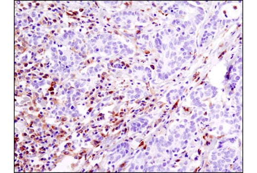
Immunohistochemical analysis of paraffin-embedded human squamous cell lung carcinoma using Btk (D3H5) Rabbit mAb performed on the Leica® Bond™ Rx.
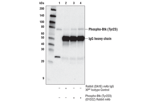
Immunoprecipitation of Phospho-Btk (Tyr223) from Ramos cells, serum-starved followed by treatment with anti-human IgM (12 μg/ml, 10 min) using Phospho-Btk (Tyr223) (D1D2Z) Rabbit mAb. Lane 1 is 10% input, lane 2 is Rabbit (DA1E) mAb IgG XP® Isotyope Control #3900, lane 3 is Phospho-Btk (Tyr223) (D1D2Z) Rabbit mAb (1:50), and lane 4 is Phospho-Btk (Tyr223) (D1D2Z) Rabbit mAb (1:100). Western blot analysis was performed using Phospho-Btk (Tyr223) (D1D2Z) Rabbit mAb. Anti-rabbit IgG, HRP-linked Antibody #7074 was used as the secondary antibody.
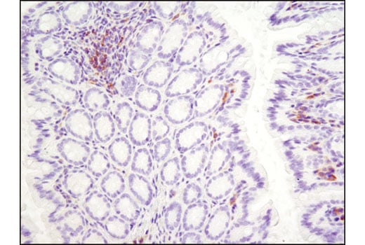
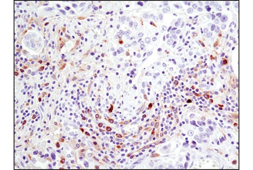
Immunohistochemical analysis of paraffin-embedded human colon carcinoma using Btk (D3H5) Rabbit mAb. Note staining of inflammatory cells.
Orders: 877-616-CELL (2355) • [email protected] • Support: 877-678-TECH (8324) • [email protected] •
Web:
cellsignal.com For Research Use Only. Not for Use in Diagnostic Procedures.
Revision 1
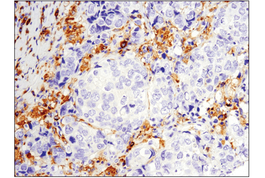
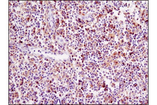
Immunohistochemical analysis of paraffin-embedded human B-cell lymphoma using Btk (D3H5) Rabbit mAb.
Immunohistochemical analysis of paraffin-embedded mouse colon using Btk (D3H5) Rabbit mAb.
Immunohistochemical analysis of paraffin-embedded human ovarian carcinoma using Btk (D3H5) Rabbit mAb. Note staining of inflammatory cells.
Orders: 877-616-CELL (2355) • [email protected] • Support: 877-678-TECH (8324) • [email protected] •
Web:
cellsignal.com For Research Use Only. Not for Use in Diagnostic Procedures.
Revision 1
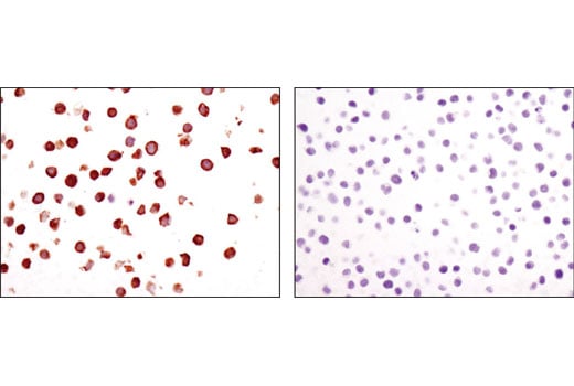
Immunohistochemical analysis of paraffin-embedded cell pellets, Ramos(left) or Jurkat (right), using Btk (D3H5) Rabbit mAb.
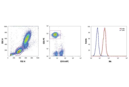
Human whole blood was fixed, lysed, and permeabilized as per the Cell Signaling Technology Flow Alternate Protocol and stained using Btk (D3H5) Rabbit mAb. Samples were co-stained using CD3-PE and CD19-APC to distinguish T and B cell subpopulations, respectively. B (red) and T (blue) cell population gates were applied to a histogram depicting the mean fluorescence intensity of Btk. Anti-rabbit IgG (H+L), F(ab')2 Fragment (Alexa Fluor® 488 Conjugate) #4412 was used as a secondary antibody.
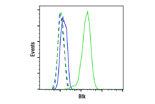
Flow cytometric analysis of Daudi cells (green) and Jurkat cells (blue) using Btk (D3H5) Rabbit mAb (solid lines) or a concentration-matched Rabbit (DA1E) mAb IgG XP® Isotype Control #3900 (dashed lines). Anti-rabbit IgG (H+L), F(ab’)2 Fragment (Alexa Fluor® 488 Conjugate) #4412 was used as a secondary antibody.
Orders: 877-616-CELL (2355) • [email protected] • Support: 877-678-TECH (8324) • [email protected] •
Web:
cellsignal.com For Research Use Only. Not for Use in Diagnostic Procedures.