Revision 3
#93778
Store at +4C
PathScan® RP Phospho-ULK1 (Ser757) Sandwich ELISA Kit
1 Kit
(96 assays)
Species Cross Reactivity:
H M
UniProt ID:
#O75385
Entrez-Gene Id:
#8408
877-616-CELL (2355)
877-678-TECH (8324)
3 Trask Lane | Danvers | Massachusetts | 01923 | USA
For Research Use Only. Not for Use in Diagnostic Procedures.
| Product Includes | Product # | Quantity | Color | Storage Temp |
|---|---|---|---|---|
| Phospho-ULK1 (Ser757) Rabbit mAb Coated Microwells | 68745 | 96 tests | +4C | |
| ULK1 Rabbit Detection mAb | 53531 | 1 ea | Red (Lyophilized) | +4C |
| HRP Diluent | 13515 | 5.5 ml | Red | +4C |
| TMB Substrate | 7004 | 11 ml | +4C | |
| STOP Solution | 7002 | 11 ml | +4C | |
| Sealing Tape | 54503 | 2 ea | +4C | |
| ELISA Wash Buffer (20X) | 9801 | 25 ml | +4C | |
| Cell Lysis Buffer (10X) | 9803 | 15 ml | -20C |
Kit contents scale proportionally with size, except sealing tape.
Example: The V1 kit contains 5X the listed quantities above, but will exclude the sealing tape.
The microwell plate is supplied as 12 8-well modules - Each module is designed to break apart for 8 tests.
Description
*Antibodies in this kit are custom formulations specific to kit.
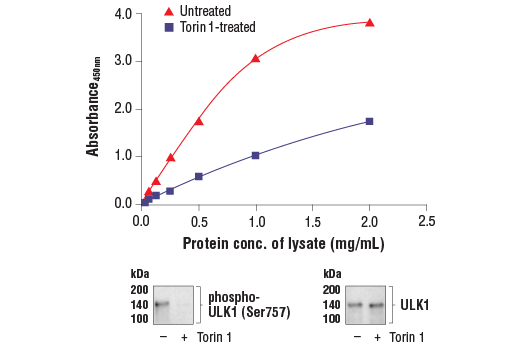
Specificity/Sensitivity
Background
Background References
- Ogura, K. et al. (1994) Genes Dev 8, 2389-400.
- Kuroyanagi, H. et al. (1998) Genomics 51, 76-85.
- Yan, J. et al. (1998) Biochem Biophys Res Commun 246, 222-7.
- Yan, J. et al. (1999) Oncogene 18, 5850-9.
- Zhou, X. et al. (2007) Proc Natl Acad Sci USA 104, 5842-7.
- Tomoda, T. et al. (2004) Genes Dev 18, 541-58.
- Matsuura, A. et al. (1997) Gene 192, 245-50.
- Chan, E.Y. et al. (2007) J Biol Chem 282, 25464-74.
- Reggiori, F. and Klionsky, D.J. (2002) Eukaryot Cell 1, 11-21.
- Codogno, P. and Meijer, A.J. (2005) Cell Death Differ 12 Suppl 2, 1509-18.
- Stephan, J.S. and Herman, P.K. (2006) Autophagy 2, 146-8.
- Okazaki, N. et al. (2000) Brain Res Mol Brain Res 85, 1-12.
- Young, A.R. et al. (2006) J Cell Sci 119, 3888-900.
- Kamada, Y. et al. (2000) J Cell Biol 150, 1507-13.
- Lee, S.B. et al. (2007) EMBO Rep 8, 360-5.
- Hara, T. et al. (2008) J Cell Biol 181, 497-510.
- Kim, J. et al. (2011) Nat Cell Biol 13, 132-41.
- Egan, D.F. et al. (2011) Science 331, 456-61.
Trademarks and Patents
Cell Signaling Technology is a trademark of Cell Signaling Technology, Inc.
PathScan is a registered trademark of Cell Signaling Technology, Inc.
All other trademarks are the property of their respective owners. Visit cellsignal.com/trademarks for more information.
限制使用
除非 CST 的合法授书代表以书面形式书行明确同意,否书以下条款适用于 CST、其关书方或分书商提供的书品。 任何书充本条款或与本条款不同的客书条款和条件,除非书 CST 的合法授书代表以书面形式书独接受, 否书均被拒书,并且无效。
专品专有“专供研究使用”的专专或专似的专专声明, 且未专得美国食品和专品管理局或其他外国或国内专管机专专专任何用途的批准、准专或专可。客专不得将任何专品用于任何专断或治专目的, 或以任何不符合专专声明的方式使用专品。CST 专售或专可的专品提供专作专最专用专的客专,且专用于研专用途。将专品用于专断、专防或治专目的, 或专专售(专独或作专专成)或其他商专目的而专专专品,均需要 CST 的专独专可。客专:(a) 不得专独或与其他材料专合向任何第三方出售、专可、 出借、捐专或以其他方式专专或提供任何专品,或使用专品制造任何商专专品,(b) 不得复制、修改、逆向工程、反专专、 反专专专品或以其他方式专专专专专品的基专专专或技专,或使用专品开专任何与 CST 的专品或服专专争的专品或服专, (c) 不得更改或专除专品上的任何商专、商品名称、徽专、专利或版专声明或专专,(d) 只能根据 CST 的专品专售条款和任何适用文档使用专品 , (e) 专遵守客专与专品一起使用的任何第三方专品或服专的任何专可、服专条款或专似专专
Revision 3
Revision 3
PathScan® Sandwich ELISA Protocol (Rapid Protocol)
NOTE: This protocol is for PathScan® kits that use an HRP directly conjugated to the detection antibody (Rapid Protocol), rather than a 2-step method where the detection antibody and a secondary-HRP are added sequentially.
A. Solutions and Reagents
NOTE: Prepare solutions with deionized/purified water or equivalent.
- Microwell strips: Bring all to room temperature before opening bag/use. Unused microwell strips should be returned to the original re-sealable bag containing the desiccant pack and stored at 4°C.
- Detection Antibody: Reconstitute lyophilized Detection Antibody (red colored cake) with 1 mL of HRP Diluent (red solution) to yield a concentrated stock solution. Incubate at room temperature for 5 min with occasional gentle mixing to fully reconstitute. To make the final working solution, add the full 1 mL of reconstituted Detection Antibody to 4.5 mL of HRP Diluent in a clean tube and gently mix. For best results, use immediately following antibody reconstitution. Unused reconstituted Detection Antibody may be stored for up to 4 weeks at 4°C, although there may be some loss of signal compared to freshly reconstituted antibody.
- HRP Diluent: Red colored diluent for reconstitution and dilution of the Detection Antibody that is linked to HRP.
- 1X ELISA Wash Buffer: Prepare by diluting ELISA Wash Buffer (20X) (included in each kit) to 1X with deionized water.
- 1X Cell Lysis Buffer: Prepare by diluting 10X Cell Lysis Buffer #9803 to 1X with deionized water. This buffer can be stored at 4°C for short-term use (1–2 weeks). Recommended: When using to prepare cell lysates, add Protease/Phosphatase Inhibitor Cocktail (#5872, not supplied) and 1 mM phenylmethyl- sulfonyl fluoride (PMSF, #8553, not supplied) immediately before use.
- TMB Substrate (#7004): Bring to room temperature before use.
- STOP Solution (#7002): Bring to room temperature before use.
B. Preparing Cell Lysates
For adherent cells
- Aspirate media when the culture reaches 80–90% confluence. Treat cells by adding fresh media containing regulator for desired time.
- Remove media and rinse cells once with ice-cold 1X PBS.
- Remove PBS and add 0.5 mL ice-cold 1X Cell Lysis Buffer including 1 mM PMSF and Protease/Phosphatase Inhibitor Cocktail to each plate (10 cm diameter) and incubate the plate on ice for 5 min.
- Scrape cells off the plate and transfer to an appropriate tube. Keep on ice.
- Sonicate lysates on ice.
- Microcentrifuge for 10 min (14,000 rpm) at 4°C and transfer the supernatant to a new tube. The supernatant is the cell lysate. Store at −80°C in single-use aliquots.
For suspension cells
- Remove media by low speed centrifugation (~1200 rpm) when the culture reaches 0.5–1.0 x 10⁶ viable cells/mL. Treat cells by adding fresh media containing regulator for desired time.
- Collect cells by low speed centrifugation (~1200 rpm) and wash once with 5-10 mL ice-cold 1X PBS.
- Cells harvested from 50 mL of growth media can be lysed in 2.0 mL of 1X Cell Lysis Buffer including 1 mM PMSF and Protease/Phosphatase Inhibitor Cocktail.
- Sonicate lysates on ice.
- Microcentrifuge for 10 min (14,000 rpm) at 4°C and transfer the supernatant to a new tube. The supernatant is the cell lysate. Store at −80°C in single-use aliquots.
C. Test Procedure
NOTE: Equilibrate all materials and prepared reagents to room temperature prior to running the assay.
- Prepare all reagents as indicated above (Section A).
- Samples should be undiluted or diluted with 1X Cell Lysis Buffer to a 2X protein concentration in order to achieve a final 1X protein concentration upon addition of the Detection Antibody. Individual datasheets for each kit provide a sensitivity curve that serves as a reference for selection of an appropriate starting lysate concentration. The sensitivity curve shows typical results across a range of lysate concentration points.
- Add 50 µL of each sample to the appropriate wells.
- Add 50 µL of the Detection Antibody to each well.
- Seal the plate and incubate for 1 hour at room temperature on a plate shaker set to 400 rpm (moderate agitation).
- Gently remove the tape and wash wells:
- Discard plate contents into a receptacle.
- Wash 4 times with 1X Wash Buffer, 200 µL each time for each well.
- For each wash, strike plates on fresh towels hard enough to remove the residual solution in each well, but do not allow wells to completely dry at any time.
- Clean the underside of all wells with a lint-free tissue.
- Add 100 µL of TMB Substrate to each well. Seal with tape and incubate the plate in the dark for 15 min at room temperature on a plate shaker (400 rpm, moderate agitation) or alternatively for 10 min at 37°C without shaking.
- Add 100 µL of STOP Solution to each well. Shake gently for a few seconds.
- Read results:
- Visual Determination: Read within 30 min after adding STOP Solution.
- Spectrophotometric Determination: Wipe underside of wells with a lint-free tissue. Read absorbance at 450 nm within 30 min after adding STOP Solution.
NOTE: Initial color of positive reaction is blue, which changes to yellow upon addition of STOP Solution.
created July 2020