Revision 4
#7036
Store at +4C
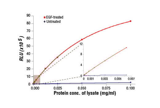
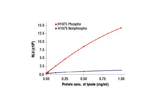
PathScan® Phospho-EGF Receptor (Tyr1068) Chemiluminescent Sandwich ELISA Kit
1 Kit
(96 assays)
Species Cross Reactivity:
H
UniProt ID:
#P00533
Entrez-Gene Id:
#1956
877-616-CELL (2355)
877-678-TECH (8324)
3 Trask Lane | Danvers | Massachusetts | 01923 | USA
For Research Use Only. Not for Use in Diagnostic Procedures.
| Product Includes | Product # | Quantity | Color | Storage Temp |
|---|---|---|---|---|
| EGF Receptor Mouse mAb Coated Microwells | 99592 | 96 tests | +4C | |
| Phospho-EGF Receptor (Tyr1068) Rabbit Detection mAb | 13019 | 1 ea | Green (Lyophilized) | +4C |
| Anti-rabbit IgG, HRP-linked Antibody (ELISA Formulated) | 13272 | 1 ea | Red (Lyophilized) | +4C |
| Detection Antibody Diluent | 13339 | 5.5 ml | Green | +4C |
| HRP Diluent | 13515 | 5.5 ml | Red | +4C |
| Luminol/Enhancer Solution | 84850 | 3 ml | RT | |
| Stable Peroxide Buffer | 42552 | 3 ml | RT | |
| Sealing Tape | 54503 | 2 ea | +4C | |
| ELISA Wash Buffer (20X) | 9801 | 25 ml | +4C | |
| ELISA Sample Diluent | 11083 | 25 ml | Blue | +4C |
| Cell Lysis Buffer (10X) | 9803 | 15 ml | -20C |
Kit contents scale proportionally with size, except sealing tape.
Example: The V1 kit contains 5X the listed quantities above, but will exclude the sealing tape.
The microwell plate is supplied as 12 8-well modules - Each module is designed to break apart for 8 tests.
Description
*Antibodies in this kit are custom formulations specific to kit.
Specificity/Sensitivity
Background
Background References
- Hackel, P.O. et al. (1999) Curr Opin Cell Biol 11, 184-9.
- Zwick, E. et al. (1999) Trends Pharmacol Sci 20, 408-12.
- Cooper, J.A. and Howell, B. (1993) Cell 73, 1051-4.
- Hubbard, S.R. et al. (1994) Nature 372, 746-54.
- Biscardi, J.S. et al. (1999) J Biol Chem 274, 8335-43.
- Emlet, D.R. et al. (1997) J Biol Chem 272, 4079-86.
- Levkowitz, G. et al. (1999) Mol Cell 4, 1029-40.
- Ettenberg, S.A. et al. (1999) Oncogene 18, 1855-66.
- Rojas, M. et al. (1996) J Biol Chem 271, 27456-61.
- Feinmesser, R.L. et al. (1999) J Biol Chem 274, 16168-73.
Trademarks and Patents
Cell Signaling Technology is a trademark of Cell Signaling Technology, Inc.
PathScan is a registered trademark of Cell Signaling Technology, Inc.
All other trademarks are the property of their respective owners. Visit cellsignal.com/trademarks for more information.
限制使用
除非 CST 的合法授书代表以书面形式书行明确同意,否书以下条款适用于 CST、其关书方或分书商提供的书品。 任何书充本条款或与本条款不同的客书条款和条件,除非书 CST 的合法授书代表以书面形式书独接受, 否书均被拒书,并且无效。
专品专有“专供研究使用”的专专或专似的专专声明, 且未专得美国食品和专品管理局或其他外国或国内专管机专专专任何用途的批准、准专或专可。客专不得将任何专品用于任何专断或治专目的, 或以任何不符合专专声明的方式使用专品。CST 专售或专可的专品提供专作专最专用专的客专,且专用于研专用途。将专品用于专断、专防或治专目的, 或专专售(专独或作专专成)或其他商专目的而专专专品,均需要 CST 的专独专可。客专:(a) 不得专独或与其他材料专合向任何第三方出售、专可、 出借、捐专或以其他方式专专或提供任何专品,或使用专品制造任何商专专品,(b) 不得复制、修改、逆向工程、反专专、 反专专专品或以其他方式专专专专专品的基专专专或技专,或使用专品开专任何与 CST 的专品或服专专争的专品或服专, (c) 不得更改或专除专品上的任何商专、商品名称、徽专、专利或版专声明或专专,(d) 只能根据 CST 的专品专售条款和任何适用文档使用专品 , (e) 专遵守客专与专品一起使用的任何第三方专品或服专的任何专可、服专条款或专似专专
Revision 4
Revision 4
ELISA Chemiluminescent (Lyophilized)
NOTE: Refer to product-specific datasheets for assay incubation temperature. This chemiluminescent ELISA is offered in low volume microplates. Only 50 µl of samples or reagents are required in each microwell.
A. Solutions and Reagents
NOTE: Prepare solutions with purified water.
- Microwell strips: Bring all to room temperature before use.
- Detection Antibody: Supplied lyophilized as a green colored cake or powder. Add 0.5 ml of Detection Antibody Diluent (green solution) to yield a concentrated stock solution. Incubate at room temperature for 5 min with occasional gentle mixing to fully reconstitute. To make the final working solution, add the 0.5 ml volume of reconstituted Detection Antibody to 5.0 ml of Detection Antibody Diluent in a clean tube and gently mix. Unused working solution may be stored for 4 weeks at 4°C.
- HRP-Linked Antibody*: Supplied lyophilized as a red colored cake or powder. Add 0.5 ml of HRP Diluent (red solution) to yield a concentrated stock solution. Incubate at room temperature for 5 min with occasional gentle mixing to fully reconstitute. To make the final working solution, add the 0.5 ml volume of reconstituted HRP-Linked Antibody to 5.0 ml of HRP Diluent in a clean tube and gently mix. Unused working solution may be stored for 4 weeks at 4°C.
- Detection Antibody Diluent: Green colored diluent for reconstitution and dilution of the detection antibody.
- HRP Diluent: Red colored diluent for reconstitution and dilution of the HRP‑Linked Antibody.
- Sample Diluent: Blue colored diluent for dilution of cell lysates.
- 1X Wash Buffer: Prepare by diluting 20X Wash Buffer (included in each PathScan® Sandwich ELISA Kit) in purified water.
- Cell Lysis Buffer: 10X Cell Lysis Buffer #9803: This buffer can be stored at 4°C for short-term use (1–2 weeks). Recommended: Add 1 mM phenylmethylsulfonyl fluoride (PMSF) immediately before use.
- Luminol/Enhancer Solution and Stable Peroxide Buffer.
*NOTE: Some PathScan® ELISA Kits may include HRP-Linked Streptavidin in place of HRP-Linked Antibody.
B. Preparing Cell Lysates
For adherent cells.
- Aspirate media when the culture reaches 80–90% confluence. Treat cells by adding fresh media containing regulator for desired time.
- Remove media and rinse cells once with ice-cold 1X PBS.
- Remove PBS and add 0.5 ml ice-cold 1X Cell Lysis Buffer plus 1 mM PMSF to each plate (10 cm diameter) and incubate the plate on ice for 5 min.
- Scrape cells off the plate and transfer to an appropriate tube. Keep on ice.
- Sonicate lysates on ice.
- Microcentrifuge for 10 min (x14,000 rpm) at 4°C and transfer the supernatant to a new tube. The supernatant is the cell lysate. Store at −80°C in single-use aliquots.
For suspension cells
- Remove media by low speed centrifugation (~1200 rpm) when the culture reaches 0.5–1.0 x 10⁶ viable cells/ml. Treat cells by adding fresh media containing regulator for desired time.
- Collect cells by low speed centrifugation (~1200 rpm) and wash once with 5–10 ml ice-cold 1X PBS.
- Cells harvested from 50 ml of growth media can be lysed in 2.0 ml of 1X Cell Lysis Buffer plus 1 mM PMSF.
- Sonicate lysates on ice.
- Microcentrifuge for 10 min (x14,000 rpm) at 4°C and transfer the supernatant to a new tube. The supernatant is the cell lysate. Store at −80°C in single-use aliquots.
C. Test Procedure
- After the microwell strips have reached room temperature, break off the required number of microwells. Place the microwells in the strip holder. Unused microwells must be resealed and stored at 4°C immediately.
- Cell lysates can be undiluted or diluted with Sample Diluent (supplied in each PathScan® Sandwich ELISA Kit, blue color). Individual datasheets for each kit provide a sensitivity curve that serves as a reference for selection of an appropriate starting lysate concentration. The sensitivity curve shows typical kit assay results across a range of lysate concentration points.
- Add 50 µl of each undiluted or diluted cell lysate to the appropriate well. Seal with tape and press firmly onto top of microwells. Incubate the plate for 2 hr at room temperature. Alternatively, the plate can be incubated overnight at 4°C.
- Gently remove the tape and wash wells:
- Discard plate contents into a receptacle.
- Wash 4 times with 1X Wash Buffer, 150 µl each time for each well.
- For each wash, strike plates on fresh towels hard enough to remove the residual solution in each well, but do not allow wells to completely dry at any time.
- Clean the underside of all wells with a lint-free tissue.
- Add 50 µl of reconstituted Detection Antibody (green color) to each well (refer to Section A, Step 2). Seal with tape and incubate the plate at room temperature for 1 hr.
- Repeat wash procedure (Section C, Step 4).
- Add 50 µl of reconstituted HRP-linked secondary antibody (red color) to each well (refer to Section A, Step 3). Seal with tape and incubate the plate at room temperature for 30 min.
- Repeat wash procedure (Section C, Step 4).
- Prepare Working Solution by mixing equal parts Luminol/Enhancer Solution and Stable Peroxide Buffer.
- Add 50 µl of the Working Solution to each well.
- Use a plate-based luminometer to measure Relative Light Units (RLU) at 425 nm within 1–10 min following addition of the substrate. Optimal signal intensity is achieved when read within 10 min.
posted November 2013
revised July 2024